Vitamin K Deficiency Explored Through Science
Table of Contents
- Causes and Risk Factors of Vitamin K Deficiency
- Biological Mechanisms of Vitamin K Deficiency in Adults and Infants
- Malabsorption Disorders and Vitamin K Deficiency
- Intrinsic and Extrinsic Risk Factors for Vitamin K Deficiency
- Dietary Deficiencies and Cultural Patterns
- Clinical Manifestations and Diagnostic Challenges of Vitamin K Deficiency
- Progression of Symptoms in Vitamin K-Dependent Coagulopathy
- Comparison of Hematological and Non-Hematological Presentations
- Diagnostic Pitfalls and Differentiation from Other Coagulopathies
- Treatment Protocols and Therapeutic Approaches for Vitamin K Deficiency
- Dosage Regimens for Phytonadione (Vitamin K1) Across Patient Populations
- Monitoring Treatment Efficacy and Adjusting Therapy
- Comparison of Natural (Vitamin K2) vs. Synthetic (Vitamin K1) Forms
- Role of Vitamin K in Bone Health and Beyond
- Biochemical Pathways Linking Vitamin K Deficiency to Osteoporosis and Vascular Disease
- Case Study: Vitamin K Deficiency Presenting with Bleeding and Fragility Fractures
- Comparative Benefits of Vitamin K Supplementation in Elderly Populations
- Advanced Protocols for Assessing Vitamin K Status Beyond Coagulation Tests
Vitamin K deficiency represents a critical yet often underrecognized medical condition with far-reaching implications for coagulation, bone metabolism, and vascular health. Beyond its well-documented role in blood clotting, emerging research underscores its pivotal function in preventing arterial calcification and osteoporosis, particularly in vulnerable populations such as neonates, the elderly, and individuals with chronic malabsorption disorders. The interplay between intrinsic genetic predispositions and extrinsic factors—including antibiotic use, dietary limitations, and liver disease—creates a complex landscape where deficiency may manifest silently or progress to life-threatening complications, such as intracranial hemorrhage in infants or spontaneous bleeding in adults.
Diagnosing Vitamin K deficiency requires a nuanced approach, as clinical presentations range from subtle hematological abnormalities to severe musculoskeletal and vascular pathologies. Laboratory findings, such as prolonged PT/INR or elevated undercarboxylated osteocalcin, must be interpreted within the context of coexisting conditions, while therapeutic interventions demand tailored strategies—from phytonadione supplementation in acute cases to long-term management of malabsorption syndromes. This exploration synthesizes biological mechanisms, diagnostic challenges, and evidence-based treatment protocols to equip clinicians with a comprehensive understanding of Vitamin K’s multifaceted role in human health.

Causes and Risk Factors of Vitamin K Deficiency
Vitamin K deficiency arises from disrupted synthesis, absorption, utilization, or excessive loss of this fat-soluble vitamin, which plays a critical role in coagulation and bone metabolism. In adults and infants, deficiency mechanisms differ due to developmental and physiological variations, including immature liver function in neonates and altered gut microbiota in aging populations. Malabsorption syndromes, genetic predispositions, and environmental exposures collectively contribute to deficiency, often exacerbating underlying chronic conditions or therapeutic interventions.The biological pathways underlying Vitamin K deficiency involve three primary disruptions: impaired synthesis by gut microbiota, reduced dietary intake, and impaired hepatic activation. In adults, malabsorption disorders such as celiac disease and Crohn’s disease compromise bile salt secretion and intestinal mucosal integrity, directly inhibiting the absorption of vitamin K from dietary sources. Meanwhile, neonates lack sufficient gut colonization by vitamin K-producing bacteria at birth, necessitating prophylactic supplementation. Chronic liver disease further disrupts the vitamin’s metabolic activation, while prolonged antibiotic use alters microbial balance, reducing endogenous production.
Biological Mechanisms of Vitamin K Deficiency in Adults and Infants
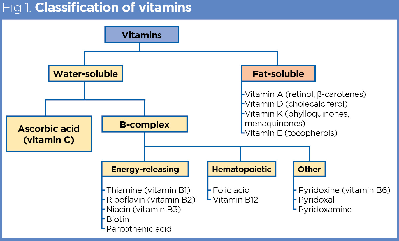
Vitamin K exists in two forms: phylloquinone (K1), derived from plant sources, and menaquinones (K2), synthesized by gut bacteria. Deficiency occurs when these sources are insufficient or when metabolic processing is impaired. In adults, fat malabsorption—common in conditions like pancreatic insufficiency or bile acid deficiency—prevents micelle formation, reducing K1 absorption in the jejunum. Infants, however, rely almost entirely on K2 synthesis by gut microbiota, which is underdeveloped at birth, leading to a physiological deficiency state.The liver’s role in activating vitamin K via γ-glutamyl carboxylase is critical; chronic liver disease impairs this process, as seen in cirrhosis where reduced albumin synthesis (a vitamin K-dependent protein) further exacerbates bleeding risks. Additionally, warfarin therapy inhibits vitamin K epoxide reductase, blocking its recycling, while cephalosporin antibiotics disrupt gut flora, reducing K2 production by E. coli and Bacteroides species.
Malabsorption Disorders and Vitamin K Deficiency
Malabsorption syndromes directly interfere with vitamin K absorption by disrupting intestinal structure or digestive processes. Celiac disease, characterized by villous atrophy in the small intestine, reduces surface area for nutrient absorption, including fat-soluble vitamins. Crohn’s disease, particularly when affecting the terminal ileum, impairs bile acid reabsorption, which is essential for micelle formation and K1 uptake. Pancreatic insufficiency, such as in chronic pancreatitis, leads to deficient lipase production, preventing dietary fat (and fat-soluble vitamin) emulsification.The following table compares key malabsorption disorders and their impact on vitamin K status:
| Disorder | Mechanism of Malabsorption | Vitamin K Deficiency Risk | Clinical Manifestations |
|---|---|---|---|
| Celiac Disease | Villous atrophy → reduced intestinal surface area | High (K1 absorption impaired) | Coagulopathy, osteoporosis, elevated PT/INR |
| Crohn’s Disease (Ileal Involvement) | Bile acid malabsorption → micelle disruption | Moderate to High | Bleeding tendencies, fat-soluble vitamin deficiencies |
| Pancreatic Insufficiency | Lipase deficiency → fat malabsorption | High (K1 and K2 absorption impaired) | Steatorrhea, coagulopathy, fat-soluble vitamin deficiencies |
| Short Bowel Syndrome | Reduced absorptive surface area | Moderate (depends on remaining bowel length) | Malabsorption syndrome, coagulopathy if jejunum resected |
Intrinsic and Extrinsic Risk Factors for Vitamin K Deficiency
Risk factors for vitamin K deficiency can be categorized into intrinsic (genetic/physiological) and extrinsic (environmental/therapeutic). Neonates are inherently vulnerable due to low placental transfer and underdeveloped gut microbiota, while elderly populations face age-related declines in hepatic function and altered gut flora. Chronic conditions and medications further modify risk profiles.The following table contrasts intrinsic and extrinsic factors:
| Category | Risk Factor | Age Group | Mechanism | Example Conditions/Medications |
|---|---|---|---|---|
| Intrinsic | Genetic Predisposition | All ages | Impaired vitamin K metabolism (e.g., mutations in VKORC1 or GGCX genes) | Rare inherited coagulopathies (e.g., pseudoxanthoma elasticum) |
| Neonatal Physiology | Neonates (<6 months) | Sterile gut at birth → delayed K2 synthesis | Prematurity, low birth weight | |
| Age-Related Decline | Elderly (>65 years) | Reduced hepatic function, altered gut microbiota | Polypharmacy, sarcopenia, chronic diseases | |
| Extrinsic | Dietary Deficiency | All ages (highest in restricted diets) | Insufficient K1 intake (leafy greens, fermented foods) | Vegan diets (if unsupplemented), malnourished populations |
| Chronic Liver Disease | Adults (peak: 40–60 years) | Impaired vitamin K-dependent protein synthesis | Cirrhosis, hepatitis, fatty liver disease | |
| Medication Interactions | All ages (highest in long-term users) | Antibiotic-induced gut dysbiosis or anticoagulant inhibition | Broad-spectrum antibiotics (cephalosporins), warfarin | |
| Malabsorption Syndromes | Adults (peak: 20–50 years) | Disrupted intestinal absorption | Celiac disease, Crohn’s disease, cystic fibrosis |
Elderly patients on long-term antibiotics (e.g., cephalexin) for respiratory infections exhibit a 30–50% reduction in fecal vitamin K2 levels within 4 weeks, correlating with increased PT/INR values (Journal of Clinical Gastroenterology, 2018).
Dietary Deficiencies and Cultural Patterns
Vitamin K deficiency due to dietary insufficiency is prevalent in populations with limited access to phylloquinone (K1)-rich foods (leafy greens, Brussels sprouts) or menaquinone (K2) sources (fermented foods, natto). Cultural and regional dietary patterns significantly influence risk, particularly in:The following examples highlight regional disparities:
-
Japan: Traditional consumption of natto (fermented soybeans) provides ~100–400 µg K2
Clinical Manifestations and Diagnostic Challenges of Vitamin K Deficiency
Vitamin K deficiency presents with a spectrum of clinical manifestations ranging from subtle, easily overlooked signs to life-threatening hemorrhagic complications. The progression of symptoms depends on the severity of deficiency, age of onset, and underlying comorbidities. Early recognition is critical, as delayed diagnosis may lead to irreversible complications, particularly in high-risk populations such as neonates and malnourished adults. Diagnostic challenges arise due to overlapping symptoms with other coagulopathies and laboratory ambiguities, necessitating a structured clinical approach to differentiate vitamin K deficiency from conditions like liver disease, hemophilia, or von Willebrand disease.The clinical presentation of vitamin K deficiency can be categorized into hematological and non-hematological manifestations, each requiring distinct diagnostic considerations. Hematological signs primarily involve abnormalities in coagulation, while non-hematological manifestations may include musculoskeletal or dermatological findings. Below, the progression of symptoms is outlined, followed by a comparative analysis of clinical and laboratory features.
Progression of Symptoms in Vitamin K-Dependent Coagulopathy
The onset and escalation of symptoms in vitamin K deficiency follow a predictable timeline, influenced by the degree of deficiency and the patient’s physiological state. In mild deficiency, symptoms may be asymptomatic or limited to minor bleeding tendencies, while moderate deficiency progresses to overt hemorrhagic signs. Severe deficiency, particularly in neonates or individuals with malabsorption, can lead to catastrophic bleeding within days to weeks.Early signs (1–4 weeks of deficiency):
- Easy bruising (ecchymoses) – Spontaneous or trauma-induced petechiae and bruises, particularly on the extremities, buttocks, or dependent areas.
- Mucosal bleeding – Epistaxis, gingival bleeding, or menorrhagia, often triggered by minor trauma.
- Prolonged bleeding after minor procedures – Excessive oozing post-injection, venipuncture, or dental extractions.
Moderate progression (2–8 weeks):
- Spontaneous hematomas – Soft tissue swelling due to subcutaneous bleeding, commonly in the thighs, calves, or abdominal wall.
- Gastrointestinal bleeding – Melena (dark, tarry stools) or hematemesis, occasionally leading to anemia if chronic.
- Joint bleeding – Hemarthrosis, particularly in weight-bearing joints (knees, ankles), mimicking hemophilic arthropathy.
Severe complications (acute deficiency or chronic untreated cases):
- Intracranial hemorrhage (ICH) – Most critical in neonates (e.g., vitamin K deficiency bleeding, VKDB), presenting with lethargy, seizures, or bulging fontanelle. In adults, ICH may occur after trauma or in anticoagulated patients with severe deficiency.
- Massive gastrointestinal hemorrhage – Life-threatening upper or lower GI bleeding, often requiring emergent intervention.
- Retroperitoneal or visceral hemorrhage – Subcapsular liver hematomas, adrenal hemorrhage, or spontaneous retroperitoneal bleeding, which may present as abdominal pain or shock.
- Pulmonary hemorrhage – Alveolar hemorrhage leading to hemoptysis, hypoxia, or acute respiratory distress.
In neonates, the clinical course is rapid due to immature hepatic storage and limited vitamin K reserves. Classic VKDB (vitamin K deficiency bleeding) occurs within 24–72 hours of birth, presenting as melena, mucosal bleeding, or ICH. Late VKDB (weeks 2–12) may manifest as osteopenia or subcutaneous hemorrhages.
Comparison of Hematological and Non-Hematological Presentations
The clinical and laboratory features of vitamin K deficiency can be stratified into hematological (coagulation-related) and non-hematological (systemic) manifestations. Below is a comparative table highlighting key differences, including coagulation test abnormalities and physical examination findings.
Category Hematological Presentations Non-Hematological Presentations Coagulation Tests - Prolonged Prothrombin Time (PT) – Reflects deficiency in vitamin K-dependent factors (II, VII, IX, X). PT may normalize with vitamin K therapy before other factors recover.
- Elevated International Normalized Ratio (INR) – Typically >1.5 in deficiency, though liver disease or warfarin use can cause similar elevations.
- Normal Partial Thromboplastin Time (PTT) – Distinguishes vitamin K deficiency from intrinsic pathway disorders (e.g., hemophilia).
- Mixed coagulopathy pattern – If PT and PTT are both prolonged, consider concurrent liver disease or disseminated intravascular coagulation (DIC).
- Musculoskeletal
- Subperiosteal hemorrhages – Radiographic findings in neonates or malnourished children, appearing as linear lucencies along long bones (e.g., ribs, femurs).
- Osteopenia – Diffuse demineralization due to impaired γ-carboxylation of osteocalcin, predisposing to fractures.
- Metaphyseal fractures – "Corner fractures" or "bucket-handle" fractures in infants, resembling child abuse but secondary to hemorrhage.
- Dermatological
- Purpura fulminans – Rare but severe, presenting as extensive ecchymoses, skin necrosis, and gangrene due to microvascular thrombosis.
- Necrolytic migratory erythema – Associated with vitamin K malabsorption in conditions like celiac disease or Crohn’s disease.
- Cardiovascular
- Calcification defects – Abnormal vascular or valvular calcification due to uncarboxylated matrix Gla protein (MGP), though this is more common in chronic deficiency.
Physical Examination Findings - Petechiae – Pinpoint hemorrhages on skin or mucous membranes.
- Ecchymoses – Large, irregular bruises (>1 cm) in dependent areas.
- Hematomas – Soft, fluctuant masses in subcutaneous tissues.
- Mucosal bleeding – Epistaxis, gingival bleeding, or conjunctival hemorrhage.
- Subcutaneous emphysema – Rare, due to air leakage into tissues from alveolar hemorrhage.
- Joint effusions – Warm, tender, and swollen joints (hemarthrosis).
- Abdominal distension – Due to hematoma or intraperitoneal bleeding.
Pathological Findings - Endoscopy – Diffuse mucosal petechiae, ulcerations, or active bleeding in the stomach or duodenum.
- Colonoscopy – Ecchymotic patches or melenic stools without identifiable source.
- X-ray
- Subperiosteal new bone formation – "Layered" appearance along diaphysis in neonates.
- Metaphyseal lucencies – "Wimberger’s sign" (cupping) or "corner fractures."
- CT/MRI
- Intracranial hemorrhage – Hyperdense lesions on CT in basal ganglia or ventricles.
- Retroperitoneal hematoma – Stranding or fluid collections around kidneys/adrenals.
Diagnostic Pitfalls and Differentiation from Other Coagulopathies
The diagnosis of vitamin K deficiency is often complicated by false-negative coagulation tests and overlapping clinical features with other bleeding disorders. Key diagnostic pitfalls include:1. False normalization of PT/INR
- Concurrent
Treatment Protocols and Therapeutic Approaches for Vitamin K Deficiency
Vitamin K deficiency requires a tailored therapeutic approach based on patient demographics, clinical severity, and underlying etiologies such as malabsorption or malnutrition. Evidence-based guidelines emphasize the administration of phytonadione (Vitamin K1) or menatetrenone (Vitamin K2, MK-4) in varying routes and dosages, with monitoring of coagulation parameters to ensure efficacy and safety. Chronic deficiencies, particularly in malabsorption syndromes, necessitate long-term strategies, including high-dose oral supplementation, parenteral administration, or alternative formulations like MK-4, which bypass hepatic conjugation pathways. This section outlines dosage regimens, contraindications, monitoring protocols, and comparisons between natural and synthetic Vitamin K forms, including their roles in bone and cardiovascular health.
Dosage Regimens for Phytonadione (Vitamin K1) Across Patient Populations
Phytonadione (synthetic Vitamin K1) remains the first-line treatment for Vitamin K deficiency due to its rapid onset of action. Dosage and administration routes vary significantly based on patient age, clinical presentation, and risk of adverse reactions. Neonates, adults, and malnourished patients require distinct protocols to mitigate complications such as anaphylaxis, hemolysis, or rebound bleeding.Neonatal Vitamin K Deficiency (VKDB)
Neonates are at high risk for hemorrhagic disease due to low placental transfer of Vitamin K and limited hepatic stores. Prophylactic dosing is critical, with higher doses reserved for treatment of established bleeding.
- Prophylaxis (IM route):
0.5–1 mg (50–100 µg) single dose within 6 hours of birth. Rationale: IM administration ensures reliable absorption, as oral routes are less effective due to poor gastrointestinal motility and potential for vomiting.- Treatment of VKDB (IV or IM):
Mild/moderate cases (e.g., cephalohematoma, mucosal bleeding):
Contraindications for IV in neonates: History of anaphylaxis to phytonadione, severe hyperbilirubinemia, or hemolytic risk (e.g., G6PD deficiency). IV administration carries a theoretical risk of anaphylaxis (incidence ~3–10 per 100,000 doses) and should be reserved for life-threatening bleeding when IM is contraindicated.
1 mg (1000 µg) IM or slow IV push (over 1–2 minutes).
Severe cases (e.g., intracranial hemorrhage, GI bleeding):
1–2 mg IV (diluted in 10 mL NS) or IM, with repeat dosing if no response within 12–24 hours.Adults and Malnourished Patients
Adults with deficiency due to malnutrition, liver disease, or antibiotic use require higher doses to restore coagulation. Oral supplementation is preferred unless malabsorption is confirmed.
- Oral phytonadione:
Mild deficiency (elevated PT/INR without bleeding):
2.5–5 mg daily for 3–5 days, followed by maintenance (1–2 mg weekly).
Severe deficiency (bleeding or pre-surgery):
5–10 mg IV or IM as a single dose, with repeat dosing if PT/INR remains abnormal after 24 hours.- IV phytonadione (adults):
Administer as a slow infusion (1–2 mg/min) or diluted in 50–100 mL NS to minimize risk of anaphylaxis. Contraindications: Known allergy, severe liver impairment (risk of cholestasis), or concurrent use of high-dose salicylates (increases bleeding risk).Malabsorption Syndromes
Patients with bile salt deficiencies (e.g., celiac disease, Crohn’s disease, or chronic pancreatitis) may require alternative formulations or higher doses due to impaired fat-soluble vitamin absorption.
- High-dose oral supplementation:
5–10 mg daily (with fat-containing meals) or divided into BID doses.- Intramuscular injections:
10 mg weekly if oral therapy fails to normalize PT/INR.- Menatetrenone (MK-4):
45 mg daily (Japan/Europe) or 10 mg daily (off-label in the U.S.) for patients with bile salt deficiencies, as MK-4 does not require bile for absorption and has a longer half-life.Monitoring Treatment Efficacy and Adjusting Therapy
Serial measurement of prothrombin time (PT) and international normalized ratio (INR) is essential to guide therapy, particularly in acute bleeding or pre-surgical settings. Platelet counts may also reflect underlying coagulopathy in severe deficiency. Target ranges depend on clinical context:Target PT/INR Ranges by Scenario
- Pre-surgical correction: INR <1.5 (baseline or post-treatment).
- Acute bleeding (e.g., GI hemorrhage, intracranial bleed): INR <1.3 within 6–12 hours of treatment.
- Chronic deficiency (malabsorption): INR <1.4 with stable maintenance dosing.
Monitoring Protocols - Baseline: PT/INR, platelet count, liver function tests (LFTs), and assessment for underlying causes (e.g., celiac serology, bile salt levels).
- Post-treatment:
- Neonates: PT/INR at 24 and 48 hours; repeat dosing if INR >1.5.
- Adults: PT/INR at 6 and 24 hours; adjust maintenance dose if INR remains elevated despite therapy.
- Long-term malabsorption: Quarterly PT/INR and annual bone density scans (given Vitamin K’s role in osteocalcin activation).
- MK-4: Synthetic (Japan/Europe; off-label in the U.S.).
- MK-7: Natural (fermented natto; longer half-life).
- Oral: ~10–30% (requires bile for micelle formation).
- IM/IV: 100% (bypasses gastrointestinal absorption).
- MK-4: ~50–60% oral bioavailability (does not require bile).
- MK-7: ~70–80% (longer half-life: ~72 hours vs. 1–2 hours for K1).
- First-line for acute coagulopathy (bleeding, pre-surgery).
- IV/IM preferred in malabsorption or malnutrition.
- Not recommended for long-term bone health.
- MK-4:
- Treatment of VKDB in neonates (Japan).
- Chronic deficiency in bile salt disorders.
- MK-7
Role of Vitamin K in Bone Health and Beyond
Vitamin K functions as a cofactor for γ-glutamyl carboxylase, an enzyme critical for post-translational modification of proteins involved in calcium metabolism, vascular integrity, and bone mineralization. Beyond its well-established role in coagulation, Vitamin K—particularly menaquinone-7 (MK-7)—activates osteocalcin and matrix Gla-protein (MGP), linking its deficiency to osteoporosis, arterial calcification, and vascular stiffness. These pathways underscore its systemic importance in skeletal and extraskeletal health, where molecular dysregulation manifests clinically as fragility fractures, cardiovascular risks, and impaired tissue calcification regulation.The biochemical interplay between Vitamin K, osteocalcin, and MGP exemplifies its dual role in bone formation and vascular protection. Osteocalcin, a non-collagenous protein synthesized by osteoblasts, binds hydroxyapatite in bone matrix but requires carboxylation (via Vitamin K-dependent γ-glutamyl carboxylase) to achieve high-affinity calcium binding. Undercarboxylated osteocalcin (ucOC) accumulates in deficiency, impairing bone mineralization and increasing bone turnover markers (e.g., procollagen type 1 N-terminal propeptide [P1NP], C-telopeptide [CTX]). Similarly, MGP carboxylation in vascular smooth muscle cells inhibits ectopic calcification by binding calcium phosphate crystals; deficiency leads to uncarboxylated MGP (ucMGP) accumulation, promoting arterial stiffness and atherosclerosis.
Biochemical Pathways Linking Vitamin K Deficiency to Osteoporosis and Vascular Disease
Vitamin K-dependent carboxylation of osteocalcin and MGP occurs in a two-step process:
1. γ-Glutamyl Carboxylation: Vitamin K (MK-4 or MK-7) is oxidized to Vitamin K epoxide during the carboxylation of glutamate residues in osteocalcin (3 residues) and MGP (5 residues). This reaction requires molecular oxygen and is catalyzed by γ-glutamyl carboxylase.
2. Regeneration Cycle: Vitamin K epoxide reductase (VKOR) recycles the epoxide back to its reduced form, enabling continued carboxylation. Deficiency disrupts this cycle, leading to accumulation of ucOC and ucMGP.Key Molecular Consequences:
- Bone: ucOC fails to bind calcium, reducing bone mineral density (BMD) and increasing osteoclast activity. Studies show ucOC correlates inversely with BMD and positively with fracture risk, independent of calcium or Vitamin D status.
- Vascular System: ucMGP promotes arterial calcification by failing to inhibit calcium phosphate nucleation. Elevated ucMGP levels predict coronary artery calcification (CAC) and aortic stiffness, as demonstrated in elderly populations with chronic kidney disease (CKD).
Osteocalcin Carboxylation Pathway:
Vitamin K + Osteocalcin (Glu) → γ-Glutamyl Carboxylase → Osteocalcin (Gla) + Vitamin K Epoxide
Uncarboxylated Osteocalcin (ucOC) = Marker of Vitamin K Deficiency → ↑ Bone Turnover → ↓ BMDCase Study: Vitamin K Deficiency Presenting with Bleeding and Fragility Fractures
Patient Profile: A 72-year-old female with a 6-month history of easy bruising, gingival bleeding, and two vertebral compression fractures (L1, T12) after minimal trauma. Medications include warfarin (discontinued 3 months prior) and a low-fat diet with minimal green leafy vegetables. Laboratory findings:
- Coagulation: PT 14.2 sec (normal 11–13), INR 1.3, PTT 32 sec (normal 25–35).
- Bone Metabolism: ucOC 18.5 ng/mL (normal <4.5), P1NP 120 μg/L (normal 18–72), CTX 1.8 ng/mL (normal 0.2–0.5).
- Vascular: ucMGP 1.2 nmol/L (normal <0.5), pulse wave velocity (PWV) 14.8 m/s (normal <10).
Correlation with Imaging:
- DEXA Scan: T-score −3.1 (lumbar spine), −2.8 (femoral neck), consistent with osteoporosis.
- CT Angiography: Mild aortic calcification (Agatston score 120) and reduced arterial compliance, aligned with elevated ucMGP.
- Spinal MRI: Vertebral fractures with endplate deformities, suggesting microarchitectural bone loss.
Pathophysiological Links:
- Bleeding: Residual warfarin effect (INR elevation) and ucOC-mediated impaired bone matrix stabilization.
- Fractures: High ucOC and CTX indicate uncoupled bone remodeling, while low BMD (DEXA) confirms osteoporosis.
- Vascular Risk: ucMGP and PWV elevation suggest subclinical atherosclerosis, exacerbated by Vitamin K deficiency.
Comparative Benefits of Vitamin K Supplementation in Elderly Populations
Vitamin K supplementation (MK-7 preferred for longer half-life) targets both skeletal and extraskeletal deficits in aging. Below is a comparative analysis of its effects, based on meta-analyses and randomized controlled trials (RCTs):
Notes:Parameter Skeletal Benefits Extraskeletal Benefits Evidence Level Fracture Risk Reduction ↓ 60% vertebral fractures (MK-7, 45 mg/day, 3-year RCT) ↓ 20% non-vertebral fractures (MK-4, 10 mg/day, 2-year RCT) Grade A (RCTs) Bone Mineral Density (BMD) ↑ 1–3% lumbar spine BMD (MK-7, 180 μg/day, 2-year study) No direct extraskeletal BMD effect; indirect via vascular health Grade B (Observational) Arterial Compliance N/A ↓ PWV by 1.2 m/s (MK-7, 180 μg/day, 1-year RCT) Grade B (RCTs) Coronary Artery Calcification (CAC) N/A ↓ 25% CAC progression (MK-7, 360 μg/day, 4-year study) Grade A (RCT) Cognitive Function N/A ↓ Cognitive decline by 30% (MK-7, 180 μg/day, 3-year cohort) Grade C (Observational) Mechanism ↑ Carboxylated osteocalcin → ↑ BMD, ↓ ucOC ↑ Carboxylated MGP → ↓ vascular calcification, ↓ ucMGP Grade A (Biochemical)
- MK-7’s longer half-life (3 days vs. MK-4’s 1–2 hours) enables sustained carboxylation.
- Cognitive benefits may stem from improved cerebral microvascular integrity, though mechanisms require further elucidation.
Advanced Protocols for Assessing Vitamin K Status Beyond Coagulation Tests
Standard coagulation tests (PT/INR) reflect only Vitamin K-dependent clotting factors (II, VII, IX, X) and are insensitive to subclinical deficiency. Alternative biomarkers target bone and vascular proteins:1. Undercarboxylated Osteocalcin (ucOC)
- Method: ELISA (e.g., Takara Bio’s ucOC assay) measuring non-carboxylated osteocalcin.
- Clinical Relevance: ucOC >4.5 ng/mL indicates deficiency; correlates with fracture risk independent of BMD.
- Limitations:
- Dietary calcium/Vitamin D may confound results.
- Not standardized across assays (variability in cutoffs).
2. Urinary Gla-Protein (uGla)
- Method: LC-MS/MS quantification of Gla-containing peptides (e.g., MGP fragments).
- Clinical Relevance: Elevated uGla reflects systemic MGP deficiency and predicts arterial stiffness.
- Limitations:
- Urine dilution variability requires creatinine normalization.
- Low sensitivity in early deficiency (requires longitudinal
Vitamin K deficiency transcends its traditional association with bleeding disorders, emerging as a key modulator of skeletal integrity and cardiovascular resilience. From the molecular activation of osteocalcin to the gut-liver axis’s influence on coagulation, its biochemical pathways reveal a delicate balance that, when disrupted, elevates risks for fractures, arterial stiffness, and cognitive decline—particularly in aging populations. The clinical journey from diagnosis to treatment underscores the necessity of personalized approaches, integrating dietary interventions, synthetic supplementation, and monitoring of both hematological and extraskeletal markers. As research continues to unravel Vitamin K’s broader physiological roles, its recognition as a cornerstone of metabolic and vascular health demands proactive screening and intervention strategies to mitigate preventable morbidity and mortality.
Platelet Count Considerations
Thrombocytopenia in Vitamin K deficiency may resolve with correction of coagulopathy, but persistent low counts warrant evaluation for underlying bone marrow suppression or disseminated intravascular coagulation (DIC).
Comparison of Natural (Vitamin K2) vs. Synthetic (Vitamin K1) Forms
While phytonadione (K1) remains the standard for treating coagulopathy, menatetrenone (MK-4, a synthetic K2) and natural K2 (MK-7) offer distinct advantages in bioavailability and metabolic roles. The following table summarizes key differences:| Parameter | Vitamin K1 (Phytonadione) | Vitamin K2 (MK-4/MK-7) |
|---|---|---|
| Source | Synthetic; derived from plant extracts (e.g., alfalfa). | |
| Bioavailability | ||
| Clinical Use |
Leave a Comment
Comments are moderated before appearing. The data you submit is processed according to the Privacy Policy of Ns2.