Vitamin B 12 Unveils Its Critical Biological and Clinical Impact

Published

Table of Contents

Vitamin B12 or cobalamin stands as a cornerstone of human metabolism, playing indispensable roles in DNA synthesis, neurological integrity, and hematopoiesis. Unlike other B vitamins, its unique cobalt-centered structure enables critical enzymatic functions, particularly in methionine synthase and methylmalonyl-CoA mutase pathways, which underpin cellular energy and neurological signaling. Deficiencies disrupt these pathways, leading to cascading effects from megaloblastic anemia to irreversible neurological damage, underscoring its irreplaceable position in health.

The bioavailability and absorption of Vitamin B12 present distinct challenges, influenced by dietary sources, intrinsic factor dependency, and metabolic interactions with folate. Animal-derived foods remain the primary natural sources, while fortified plant-based alternatives offer alternatives for vegans and vegetarians. However, malabsorption disorders and drug interactions—such as those caused by proton pump inhibitors or metformin—further complicate optimal intake. Understanding these mechanisms is essential for accurate diagnosis and targeted therapeutic interventions.

Vitamin B12: Molecular Structure, Chemical Properties, and Cofactor Mechanisms

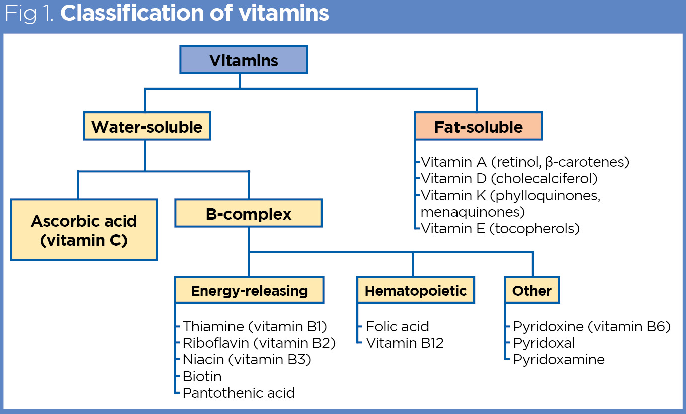
Vitamin B12, or cobalamin, is the largest and most structurally complex vitamin in human biology, featuring a corrin ring system that distinguishes it from other B vitamins. Unlike riboflavin (B2), niacin (B3), or pyridoxine (B6), which rely on simpler heterocyclic structures, B12 contains a central cobalt ion coordinated to four nitrogen atoms in a square planar arrangement, forming the corrin macrocycle. This cobalt center undergoes reversible redox cycling between Co(I), Co(II), and Co(III) oxidation states, enabling its role as a cofactor in enzymatic reactions. The unique chemical properties of B12—including its ability to stabilize organometallic intermediates and facilitate radical-based chemistry—are critical for its biological functions, which are fundamentally different from those of other B vitamins.

The biochemical versatility of B12 arises from its two primary coenzyme forms: adenosylcobalamin (AdoCbl) and methylcobalamin (MeCbl). These forms serve as essential cofactors in two distinct enzymatic pathways: methylation reactions (via methionine synthase) and isomerization reactions (via methylmalonyl-CoA mutase). The structural and electronic properties of the cobalt center allow B12 to participate in radical-mediated rearrangements, a mechanism absent in other B vitamins. Below, the molecular distinctions and enzymatic mechanisms are explored in detail.

Molecular Structure and Unique Chemical Properties of Cobalamin

The corrin ring of B12 differs from the porphyrin rings in heme or chlorophyll by replacing two of the four pyrrole rings with direct carbon-carbon bonds, reducing the aromatic stability but enhancing flexibility. This structural adaptation accommodates the upper (α) and lower (β) axial ligands, which are critical for coenzyme function. In adenosylcobalamin (AdoCbl), the cobalt is bound to 5'-deoxyadenosyl, a nucleotide derivative that undergoes homolytic cleavage to generate a 5'-deoxyadenosyl radical (5'-dA•). This radical abstracts a hydrogen atom from the substrate, initiating isomerization or rearrangement.

In contrast, methylcobalamin (MeCbl) contains a methyl group (CH₃) as the lower ligand, enabling methyl transfer in S-adenosylmethionine (SAM)-dependent reactions. The cobalt-carbon bond in MeCbl is the only known organometallic bond in mammalian biochemistry, distinguishing B12 from all other vitamins. The redox potential of the Co(III)/Co(II) couple (~−0.6 V) allows B12 to participate in single-electron transfer (SET) reactions, a feature exploited in both methionine synthase and methylmalonyl-CoA mutase.

Key Structural Features:
  • Corrin ring: Tetrapyrrole macrocycle with reduced aromaticity compared to porphyrins.
  • Cobalt center: Coordination to four nitrogen atoms in a square planar geometry, with axial ligands determining coenzyme form.
  • Organometallic bond: Unique Co-C bond in MeCbl, enabling methyl transfer.
  • Radical chemistry: AdoCbl generates 5'-dA• to facilitate substrate rearrangement.
  • Cofactor Role in Methionine Synthase and Methylmalonyl-CoA Mutase Pathways

    Vitamin B12 functions as a cofactor in two evolutionarily conserved enzymatic pathways: methionine synthase (MS) and methylmalonyl-CoA mutase (MUT). These enzymes rely on distinct chemical mechanisms enabled by the B12 coenzyme forms, highlighting the vitamin’s dual role in one-carbon metabolism and propionate catabolism.

    ### 1. Methionine Synthase (MS) Pathway: Methylation via Methylcobalamin
    Methionine synthase catalyzes the remethylation of homocysteine to methionine, a reaction essential for SAM synthesis and DNA methylation. The enzyme requires MeCbl, folate (as 5-methyltetrahydrofolate, 5-MTHF), and zinc as cofactors. The reaction proceeds via a methyl transfer cascade:

  • Step 1: 5-MTHF donates a methyl group to homocysteine, forming methionine and tetrahydrofolate (THF).
  • Step 2: The methyl group is transiently bound to the cobalt of MeCbl, forming a cob(III)alamin-methyl intermediate.
  • Step 3: The methyl group is transferred to homocysteine, regenerating hydroxocobalamin (OH-Cbl).
  • Step 4: Vitamin B12 reductase (MTRR) reduces OH-Cbl back to MeCbl using NADPH.
  • Biochemical Equation:
    5-MTHF + Homocysteine + MeCbl → THF + Methionine + OH-Cbl (regenerated to MeCbl via MTRR)
    Deficiency Impact: Impaired MS activity leads to elevated homocysteine (hyperhomocysteinemia) and folate trapping (as 5-MTHF), exacerbating neurological deficits and megablastic anemia.

    ### 2. Methylmalonyl-CoA Mutase (MUT) Pathway: Isomerization via Adenosylcobalamin
    Methylmalonyl-CoA mutase catalyzes the isomerization of L-methylmalonyl-CoA to succinyl-CoA, a critical step in branched-chain amino acid (BCAA) and odd-chain fatty acid metabolism. The reaction involves a radical rearrangement mechanism:

  • Step 1: AdoCbl cleaves homolytically to generate 5'-dA• and cob(II)alamin.
  • Step 2: The radical abstracts a hydrogen from L-methylmalonyl-CoA, forming a substrate radical intermediate.
  • Step 3: The intermediate undergoes 1,2-rearrangement, converting the substrate to succinyl-CoA.
  • Step 4: The radical is quenched by recombination with 5'-dA•, regenerating AdoCbl.
  • Biochemical Equation:
    L-Methylmalonyl-CoA + AdoCbl → Succinyl-CoA + 5'-Deoxyadenosine + Cob(II)alamin (regenerated to AdoCbl)
    Deficiency Impact: MUT deficiency results in methylmalonic aciduria (MMA), characterized by accumulation of methylmalonyl-CoA, leading to metabolic acidosis, developmental delays, and pancreatitis.

    Comparison of Vitamin B12’s Biological Roles in DNA Synthesis, Hematopoiesis, and Neurological Function

    Vitamin B12 is indispensable for three major physiological processes, each dependent on distinct metabolic pathways. Below is a comparative analysis of its roles, key enzymes, and biochemical consequences of deficiency.

    Sources and Bioavailability of Vitamin B12

    Vitamin B12, or cobalamin, is an essential nutrient primarily obtained through dietary intake, as human and plant systems lack endogenous synthesis pathways. Its bioavailability is influenced by both intrinsic and extrinsic factors, including dietary sources, gut physiology, and biochemical forms. Animal-derived foods remain the most concentrated natural sources, while fortified plant-based alternatives address deficiencies in vegan or vegetarian diets. The absorption process relies on gastric intrinsic factor (IF), a glycoprotein secreted by parietal cells, which binds B12 in the stomach and facilitates its uptake in the ileum. Disruptions in this mechanism, such as in autoimmune atrophic gastritis or pernicious anemia, lead to malabsorption and deficiency. Below, dietary sources are categorized, absorption mechanisms are detailed, and bioavailability comparisons among B12 analogs are provided alongside nutrient retention data in common foods.

    Categorized Dietary Sources of Vitamin B12

    Vitamin B12 occurs naturally in animal tissues due to microbial synthesis in the gastrointestinal tracts of ruminants and marine organisms. Plant-based sources are typically fortified to compensate for the absence of endogenous cobalamin. The following lists distinguish between natural and fortified options, with emphasis on bioavailability and practical dietary relevance.
    • Animal-Based Sources (Natural Occurrence)
      These foods contain biologically active forms of B12 (methylcobalamin or adenosylcobalamin) bound to proteins, requiring gastric digestion for release. Liver and organ meats are particularly dense in B12, followed by fish, shellfish, and dairy products.
      • Liver (beef, chicken, lamb): 50–70 µg per 100g (raw); cooking reduces content by ~10–20%.
      • Clams and oysters: 30–90 µg per 100g (raw); high bioavailability due to low protein binding.
      • Fatty fish (salmon, mackerel, trout): 5–20 µg per 100g (raw); cooking may reduce levels by 30%.
      • Eggs (yolk): 1–2 µg per 100g; pasteurization or overcooking diminishes B12 by ~50%.
      • Dairy (milk, cheese, yogurt): 0.4–1.5 µg per 100g; fermented products (e.g., Swiss cheese) retain higher levels.
      • Meat (beef, pork, poultry): 1–6 µg per 100g; lean cuts contain less than fatty or organ meats.
    • Fortified Plant-Based Sources
      Synthetic cyanocobalamin is added to plant milks, cereals, and nutritional yeast to meet dietary needs. Bioavailability varies by formulation, with fortified foods typically providing 1.2–2.5 µg per serving.
      • Nutritional yeast: 1–10 µg per 10g; active forms (methylcobalamin) may be added in premium products.
      • Fortified plant milks (soy, almond, oat): 0.6–1.2 µg per 250ml; stability decreases with light exposure.
      • Breakfast cereals: 1.5–6 µg per serving; often combined with folic acid for enhanced absorption.
      • Vegan meat substitutes: 0.3–1.5 µg per 100g; varies by manufacturer and processing.
    • Microbial Sources (Emerging Options)
      Fermented foods like tempeh (from soybeans) or miso contain trace amounts of B12 produced by bacteria, but levels are insufficient for dietary reliance. Research into Lactobacillus-enriched foods is ongoing for vegan applications.

    Mechanisms of Vitamin B12 Absorption and Malabsorption

    The absorption of Vitamin B12 is a multi-step process requiring gastric, pancreatic, and ileal factors. Intrinsic factor (IF), secreted by parietal cells in the stomach, binds free B12 released from dietary proteins via pepsin and hydrochloric acid. The IF-B12 complex then interacts with cubilin and amnionless receptors in the ileum, enabling endocytosis and transcytosis into portal circulation. Malabsorption arises from defects in any of these stages, particularly in conditions involving:
    • Gastric Atrophy or Hypochlorhydria
      Atrophic gastritis, often autoimmune (pernicious anemia), destroys parietal cells, reducing IF production. Achlorhydria (low stomach acid) further impairs protein digestion, leaving B12 bound to dietary proteins and unavailable for IF binding.
    • Ileal Dysfunction
      Crohn’s disease, celiac disease, or surgical resection of the ileum disrupt cubilin receptor-mediated uptake. Bacterial overgrowth (e.g., Serratia spp.) in the ileum competes for B12, exacerbating deficiency.
    • Transcobalamin II (TCN2) Deficiency
      Rare genetic disorders or acquired TCN2 insufficiency prevent B12 transport from enterocytes to tissues, leading to functional deficiency despite normal serum levels.
    • Drug Interactions
      Proton pump inhibitors (PPIs) and H2 blockers suppress gastric acid, reducing B12 release from food. Metformin may impair ileal absorption via unknown mechanisms.
    Physiological Consequences of Malabsorption
    Unabsorbed B12 accumulates in the gut, where bacteria metabolize it into inactive analogs (e.g., pseudovitamin B12), falsely elevating serum levels while tissues remain deficient. Neurological symptoms (e.g., peripheral neuropathy) may precede hematological signs (e.g., megaloblastic anemia) due to delayed transport of adenosylcobalamin to mitochondria.

    Bioavailability Comparisons of Vitamin B12 Analogs

    The three primary forms of Vitamin B12—cyanocobalamin, methylcobalamin, and adenosylcobalamin—differ in stability, absorption efficiency, and clinical applications. Cyanocobalamin, the most stable and cost-effective form, is widely used in supplements and fortification but requires conversion to active cofactors in the body. Methylcobalamin and adenosylcobalamin are biologically active but less stable, with distinct roles in methylation and energy metabolism, respectively.
    Bioavailability Summary:
    • Cyanocobalamin
    • Stability: High; resistant to heat, light, and oxidation. Shelf life >2 years.
    • Absorption: Requires conversion to methylcobalamin/adenosylcobalamin via hepatic enzymes. Bioavailability ~50% oral, ~90% parenteral (intramuscular/subcutaneous).
    • Suitability: Ideal for fortification and long-term supplementation. Less effective for neurological conditions due to cyanide moiety (though detoxified by rhodanese).
    • Methylcobalamin
    • Stability: Moderate; degrades faster than cyanocobalamin, especially in liquid forms. Shelf life ~1 year.
    • Absorption: Directly utilized by tissues; bioavailability ~50–100% oral (higher in deficient individuals). Preferred for methylation support (e.g., homocysteine metabolism).
    • Suitability: Optimal for neurological deficits (e.g., peripheral neuropathy), depression, and cognitive impairment. Avoid in nitrite-sensitive individuals (theoretical risk of methemoglobinemia).
    • Adenosylcobalamin
    • Stability: Lowest; highly sensitive to heat and light. Shelf life <6 months in optimal conditions.
    • Absorption: Requires conversion to methylcobalamin for transport; bioavailability ~30–50% oral. Primarily utilized in mitochondria for succinyl-CoA synthesis.
    • Suitability: Critical for energy metabolism (e.g., chronic fatigue, mitochondrial disorders). Often combined with methylcobalamin in clinical settings.
    Note: Sublingual and nasal forms of methylcobalamin bypass gastric absorption, achieving ~50–70% bioavailability without IF dependence.

    Vitamin B12 Content in Common Foods and Nutrient Retention

    Food processing methods significantly affect B12 retention, with heat, light exposure, and prolonged storage reducing bioavailability. The table below compares raw and cooked preparations, highlighting percentage losses and recommended consumption practices.
    Biological Role Key Metabolic Process Enzyme Involved Deficiency Consequence Biochemical Marker
    DNA Synthesis
    • Provides methyl groups for dTMP synthesis via thymidylate synthase (dependent on 5,10-MTHF).
    • Supports purine synthesis by maintaining folate in active forms (THF, 5-MTHF).
    • Methionine synthase (MS)
    • Thymidylate synthase (TS)
    • Impaired DNA replication → megablastic anemia.
    • Neutrophil hypersegmentation due to defective cell division.
    • ↑ Homocysteine
    • ↓ Methionine
    • ↓ 5-MTHF (folate trap)
    Red Blood Cell Production
    • Supports erythropoiesis by ensuring adequate dTMP and methionine for hemoglobin synthesis.
    • Maintains membrane integrity via S-adenosylmethionine (SAM)-dependent phospholipid methylation.

    Deficiency Symptoms and Diagnostic Approaches in Vitamin B12 Deficiency

    Vitamin B12 deficiency presents a heterogeneous clinical spectrum, ranging from subclinical biochemical abnormalities to severe, irreversible neurological and hematological complications. Early recognition relies on a structured understanding of its systemic, hematological, and neurological manifestations, alongside precise diagnostic biomarkers. Misinterpretation of laboratory results or failure to differentiate B12 deficiency from other macrocytic anemias can delay intervention, exacerbating irreversible damage. This section systematically categorizes clinical presentations, outlines diagnostic workflows, and evaluates the comparative utility of biomarkers in identifying early-stage deficiency.

    Clinical Manifestations of Vitamin B12 Deficiency

    The symptoms of vitamin B12 deficiency emerge gradually and are often nonspecific in early stages, complicating timely diagnosis. Deficiency disrupts DNA synthesis and methyl group metabolism, leading to systemic, hematological, and neurological consequences. Systemic symptoms reflect generalized metabolic dysfunction, while hematological manifestations arise from impaired erythropoiesis, and neurological complications stem from myelin degradation in the central and peripheral nervous systems.

    Systemic Symptoms
    Vitamin B12 deficiency frequently presents with nonspecific constitutional symptoms that overlap with other chronic conditions, including:

  • Fatigue and weakness, often progressive and disproportionate to physical exertion, resulting from impaired ATP production and mitochondrial dysfunction.
  • Pallor and jaundice, secondary to ineffective erythropoiesis and hemolysis, with scleral icterus and dark urine in severe cases.
  • Glossitis and angular cheilitis, reflecting mucosal atrophy and inflammation due to impaired cellular turnover.
  • Weight loss and anorexia, attributed to malabsorption or underlying gastrointestinal disorders (e.g., atrophic gastritis, celiac disease).
  • Hematological Manifestations
    The hallmark of B12 deficiency is megaloblastic anemia, characterized by:

  • Macrocytosis (MCV > 100 fL) with hypersegmented neutrophils (>5 lobes), though macrocytosis may also occur in alcoholism, liver disease, or hypothyroidism.
  • Anisopoikilocytosis, with large, oval-shaped red blood cells (macrocytes) and irregularly shaped cells (poikilocytes).
  • Reticulocytopenia, despite anemia, due to ineffective erythropoiesis in the bone marrow.
  • Elevated lactate dehydrogenase (LDH) and indirect bilirubin, indicating hemolysis from premature RBC destruction.
  • Neurological Complications
    Neurological damage is often irreversible if untreated, involving:

  • Peripheral neuropathy, presenting as numbness, tingling, or burning sensations in the hands and feet, progressing to motor weakness and loss of reflexes.
  • Subacute combined degeneration (SCD) of the spinal cord, characterized by posterior column dysfunction (vibration and proprioception loss) and corticospinal tract involvement (spasticity, hyperreflexia).
  • Cognitive decline, including memory impairment, dementia-like symptoms, and mood disturbances (depression, irritability), attributed to demyelination in the cerebral cortex.
  • Optic neuropathy, rare but severe, leading to visual disturbances or blindness due to retinal nerve fiber layer degeneration.
  • Diagnostic Biomarkers and Laboratory Interpretation

    Accurate diagnosis of vitamin B12 deficiency requires a multimodal approach, integrating serum B12 levels with functional biomarkers (methylmalonic acid [MMA] and homocysteine) to overcome limitations of isolated tests. Serum B12 assays alone are unreliable due to high false-negative rates, particularly in early deficiency or high plasma transcobalamin II (TCII) levels. Functional biomarkers reflect downstream metabolic consequences of B12 deficiency, improving diagnostic sensitivity.

    Step-by-Step Interpretation of Laboratory Results
    The diagnostic workflow follows a hierarchical approach to minimize false positives/negatives:

    1. Initial Screening: Serum Vitamin B12

  • Reference range: 200–900 pg/mL (varies by assay; some labs use 250–600 pg/mL).
  • Deficiency thresholds:
  • <200 pg/mL: Strongly suggestive of deficiency, though functional biomarkers should be confirmed.
  • 200–400 pg/mL: "Borderline" range; functional tests (MMA/homocysteine) are mandatory.
  • >400 pg/mL: Typically excludes deficiency, except in cases of functional B12 deficiency (e.g., genetic TCII mutations or malabsorption with elevated "trapped" B12 in tissues).
  • Limitations: Up to 50% of deficient patients may have normal serum B12 due to assay interference (e.g., high haptocorrin levels) or tissue-bound B12.
  • 2. Functional Biomarkers: Methylmalonic Acid (MMA) and Homocysteine

  • MMA (primary marker for B12 deficiency):
  • Reference range: 73–271 nmol/L (varies by age; higher in elderly).
  • Deficiency threshold: >271 nmol/L (or >400 nmol/L in some labs) indicates B12-related metabolic dysfunction.
  • Specificity: Elevated MMA is specific to B12 deficiency (not folate deficiency), as MMA accumulation results from impaired methylmalonyl-CoA mutase activity.
  • Homocysteine (secondary marker, reflects B12/folate status):
  • Reference range: 4–14 µmol/L.
  • Deficiency threshold: >14 µmol/L (elevated in both B12 and folate deficiency).
  • Limitations: Homocysteine elevation is non-specific; it also rises in renal disease, hypothyroidism, or drug-induced deficiencies (e.g., methotrexate).
  • 3. Confirmatory Tests for Malabsorption

  • Schilling Test (historical, rarely used): Assesses oral B12 absorption with/without intrinsic factor; replaced by intrinsic factor antibody (IFA) testing and gastric analysis (e.g., serum gastrin, pepsinogen I).
  • Intrinsic Factor Antibodies (IFA): Positive in pernicious anemia (autoimmune atrophic gastritis).
  • Parietal Cell Antibodies (PCA): Supports diagnosis of autoimmune gastritis.
  • Gastric Biopsy: Confirms atrophic gastritis or Helicobacter pylori infection in malabsorptive cases.
  • Diagnostic Algorithm for Biomarker Comparison
    The following table summarizes the diagnostic accuracy of biomarkers in early-stage deficiency:

    BiomarkerSensitivitySpecificityFalse-Negative ScenariosFalse-Positive Scenarios
    Serum B12Low (30–50%)ModerateEarly deficiency, high haptocorrin, TCII mutationsPregnancy, liver disease, high transcobalamin I
    MMAHigh (80–90%)HighRenal impairment (MMA clearance)Rare (e.g., genetic MMA mutase deficiency)
    HomocysteineModerate (60%)LowFolate deficiency, renal disease, hypothyroidismB6 deficiency, estrogen therapy
    Key Insight:
  • MMA is the gold standard for early B12 deficiency, particularly in patients with normal serum B12 but clinical suspicion (e.g., neuropathy or cognitive symptoms).
  • Homocysteine elevation alone cannot distinguish B12 from folate deficiency; MMA must be checked.
  • Combined MMA + homocysteine testing improves sensitivity to ~95% for B12 deficiency.
  • Differentiating Vitamin B12 Deficiency from Other Causes of Macrocytic Anemia

    Macrocytic anemia encompasses a broad differential diagnosis, requiring systematic exclusion of alternative etiologies. Vitamin B12 deficiency must be distinguished from folate deficiency, alcohol-related macrocytosis, hypothyroidism, liver disease, and drug-induced megaloblastosis. A flowchart-based approach ensures logical exclusion of these conditions.

    Flowchart Decision-Making Process
    1. Step 1: Confirm Macrocytosis (MCV > 100 fL)

  • If MCV < 100 fL, exclude B12 deficiency; consider iron deficiency or thalassemia.
  • 2. Step 2: Assess Reticulocyte Count

  • Reticulocytopenia (<2%): Suggests ineffective erythropoiesis (B12/folate deficiency, myelodysplasia).
  • Reticulocytosis (>2%): Indicates hemolytic anemia (e.g., cold agglutinin disease) or recovery from aplasia.
  • 3. Step 3: Evaluate Functional Biomarkers

  • MMA elevated + Homocysteine elevated: B12 deficiency (proceed to intrinsic
  • Vitamin B12 in Health and Disease: Cardiovascular, Neurological, and Pharmacological Interactions

    Vitamin B12 plays a critical role in maintaining cardiovascular and neurological health, with its deficiency linked to systemic dysfunctions ranging from endothelial damage to neurodegenerative progression. Epidemiological evidence underscores its involvement in homocysteine metabolism, mitochondrial integrity, and inflammatory pathways, while drug-nutrient interactions further complicate its therapeutic optimization. This section examines the mechanistic links between B12 status and cardiovascular risks, its potential role in neurodegenerative diseases, clinical supplementation guidelines for vulnerable populations, and pharmacodynamic interactions with common medications.

    Cardiovascular Implications of Vitamin B12: Homocysteine, Endothelial Function, and Epidemiological Evidence

    Vitamin B12 functions as a cofactor in the remethylation of homocysteine to methionine, a reaction catalyzed by methionine synthase (MS). Elevated homocysteine levels, a hallmark of B12 deficiency, are independently associated with increased cardiovascular risk through oxidative stress, endothelial dysfunction, and vascular smooth muscle proliferation. Observational studies, including the Framingham Heart Study and Physicians’ Health Study, demonstrate that higher homocysteine concentrations correlate with a 2- to 3-fold increased risk of myocardial infarction and stroke, even after adjusting for traditional risk factors (e.g., hypertension, diabetes, smoking).

    The Norwegian Vitamin Trial (NORVIT), a large randomized controlled trial (RCT), investigated the effects of B12 and folate supplementation on secondary cardiovascular prevention. While the study found no significant reduction in major adverse cardiovascular events (MACE) in the general population, subgroup analyses revealed modest benefits in patients with preexisting hyperhomocysteinemia (baseline homocysteine >13 µmol/L). Mechanistically, B12 deficiency impairs nitric oxide (NO) bioavailability by reducing tetrahydrobiopterin (BH4) synthesis, a cofactor for endothelial nitric oxide synthase (eNOS). This leads to endothelial dysfunction, characterized by reduced vasodilation and increased platelet aggregation. A meta-analysis of 27 studies (2019) confirmed that B12 supplementation (1–2 mg/day) significantly lowered homocysteine levels by 25–30% in deficient individuals, though its direct impact on cardiovascular outcomes remains debated due to trial heterogeneity.

    Emerging evidence suggests that chronic B12 deficiency contributes to neurodegenerative pathologies through mitochondrial dysfunction, neuroinflammation, and epigenetic dysregulation. The brain’s high metabolic demand makes it particularly susceptible to B12-related disruptions in methylation cycles (e.g., SAMe synthesis) and succinyl-CoA metabolism (via methylmalonyl-CoA mutase), leading to neuroaxonal degeneration and white matter lesions. Key associations include:

    - Alzheimer’s Disease (AD):
    Postmortem studies reveal B12 deficiency in 30–50% of AD patients, with elevated methylmalonic acid (MMA) and homocysteine levels correlating with cognitive decline and hippocampal atrophy. The "vascular hypothesis" proposes that B12 deficiency exacerbates cerebral hypoperfusion by promoting amyloid-β aggregation and tau hyperphosphorylation, though direct causality requires further longitudinal studies.

    - Multiple Sclerosis (MS):
    Observational data from the US National Health and Nutrition Examination Survey (NHANES) indicate that low B12 status is associated with a 40% higher risk of MS, potentially via mitochondrial DNA damage and oligodendrocyte dysfunction. Animal models demonstrate that B12 supplementation reduces demyelination in experimental autoimmune encephalomyelitis (EAE), a rodent MS analog, by modulating microRNA-155 and interferon-γ pathways.

    - Neuropsychiatric Disorders:
    Deficiency-induced hyperhomocysteinemia may contribute to depression and cognitive impairment by disrupting serotonin and dopamine synthesis. A 2021 cohort study in JAMA Neurology found that elderly patients with subclinical B12 deficiency (serum B12 <300 pg/mL) exhibited accelerated brain volume loss in frontal and temporal lobes, independent of vitamin D or folate levels.

    Proposed Mechanisms:

    1. Mitochondrial Dysfunction: B12-dependent enzymes (e.g., methylmalonyl-CoA mutase) are critical for energy metabolism; their impairment leads to oxidative stress and neuronal apoptosis.
    2. Epigenetic Alterations: Reduced SAMe availability disrupts DNA methylation, affecting genes linked to neurogenesis (e.g., BDNF) and apoptosis (e.g., p53).
    3. Neuroinflammation: B12 deficiency elevates TNF-α and IL-6, promoting microglial activation and blood-brain barrier (BBB) permeability.
    4. Vascular Contributions: Endothelial damage (as described earlier) reduces cerebral blood flow, exacerbating hypoxic injury in vulnerable regions.

    Clinical Guidelines for Vitamin B12 Supplementation in High-Risk Populations

    Targeted supplementation is essential for populations at elevated risk of deficiency due to malabsorption, increased demand, or reduced dietary intake. The following table summarizes evidence-based guidelines from the World Health Organization (WHO), National Institutes of Health (NIH), and European Society for Clinical Nutrition and Metabolism (ESPEN):
    Population Dosage Recommendation Monitoring Protocol Key Considerations
    Pregnant Women
    • Dietary: 2.6 µg/day (RDA, increased from 2.4 µg for non-pregnant adults).
    • Supplementation: 400–800 µg/day (oral) or 1,000 µg/month (intramuscular) for deficient individuals (serum B12 <200 pg/mL).
    • High-risk groups (e.g., vegans): 1,000 µg/day oral or weekly 1,000 µg IM.
    • Baseline: Serum B12, MMA, and homocysteine at 12–16 weeks gestation.
    • Follow-up: Repeat at 28–32 weeks if initial levels are low.
    • Neonatal screening: Cord blood B12 if maternal deficiency is suspected.
    • Deficiency linked to neural tube defects (NTDs) and low birth weight.
    • Vegan diets may require longer supplementation due to poor bioavailability.
    • Avoid excessive doses (>2,000 µg/day) without monitoring due to potential masking of pernicious anemia.
    Lactating Women
    • Dietary: 2.8 µg/day (RDA).
    • Supplementation: 500–1,000 µg/day for deficient mothers or infants with exclusive breastfeeding and maternal deficiency.
    • Maternal: Serum B12 at 6 weeks postpartum.
    • Infant: Plasma B12 at 2–4 months if breastfed exclusively.
    • Breast milk B12 concentrations are low in deficient mothers, risking infant megaloblastic anemia.
    • Formula-fed infants are less at risk but may require supplementation if maternal intake is inadequate.
    Elderly (≥65 Years)
    • General population: 2.4 µg/day (RDA), with fortified foods (e.g., cereals) encouraged.
    • Deficient (serum B12 <300 pg/mL or MMA >271 nmol/L): 500–1,000 µg/day oral or 1,000 µg IM weekly for 4–8 weeks, followed by maintenance (1,000 µg monthly).
    • Supplementation and Therapeutic Applications of Vitamin B12

      Vitamin B12 supplementation plays a critical role in correcting deficiencies, optimizing neurological and metabolic functions, and addressing conditions where endogenous synthesis or absorption is impaired. The selection of supplementation form—whether oral, sublingual, intramuscular (IM), or nasal—depends on clinical presentation, patient compliance, and underlying pathophysiology. High-dose B12 therapy has demonstrated efficacy in peripheral neuropathy, chronic fatigue syndrome, and mood disorders, though mechanisms and optimal dosing remain areas of ongoing research. This section examines the pharmacokinetics of various B12 supplementation modalities, evidence-based therapeutic applications, and comparative efficacy in long-term management.

      Forms of Vitamin B12 Supplementation and Pharmacokinetics

      Vitamin B12 supplementation is available in multiple formulations, each with distinct absorption profiles, onset of action, and clinical utility. The choice of administration route influences bioavailability, particularly in patients with malabsorption syndromes (e.g., atrophic gastritis, pernicious anemia, or ileal resection). Below are the primary forms, their absorption mechanisms, and recommended protocols for deficiency correction.

      Oral Supplementation
      Oral B12 (cyanocobalamin or methylcobalamin) is the most common form due to its accessibility and cost-effectiveness. However, absorption is limited by intrinsic factor (IF)-dependent mechanisms in the ileum, with only 1–5% of ingested B12 absorbed via passive diffusion at high doses (≥1000 µg). This makes oral supplementation less effective in patients with gastric or intestinal malabsorption, where IM administration is preferred.

      Sublingual and Buccal Administration
      Sublingual (under the tongue) or buccal (cheek) tablets bypass first-pass hepatic metabolism and achieve higher bioavailability (~30–50%) compared to oral forms. This route is particularly useful for patients with mild to moderate malabsorption who cannot tolerate injections. Studies suggest 1000–2000 µg/day of sublingual methylcobalamin can restore serum levels in deficient individuals within 4–8 weeks.

      Intramuscular (IM) Injections
      IM administration of hydroxocobalamin or cyanocobalamin provides 100% bioavailability and is the gold standard for severe deficiency or malabsorption. A typical regimen involves 1000 µg weekly for 4–8 weeks, followed by 1000 µg monthly for maintenance. IM injections are also used in acute neurological presentations (e.g., subacute combined degeneration) to rapidly elevate CNS B12 levels.

      Nasal Sprays and Transdermal Gels
      Nasal sprays (e.g., 500 µg cyanocobalamin per spray) offer an alternative for patients unable to tolerate injections or oral forms, with absorption via nasal mucosa achieving ~10–20% bioavailability. Transdermal gels (e.g., 1% methylcobalamin in a gel base) are emerging but lack standardized dosing protocols; patchy absorption limits their efficacy compared to IM routes.

      Key Considerations for Supplementation Protocols

    • Deficiency Correction: IM injections are preferred for serum B12 <200 pg/mL or elevated methylmalonic acid (MMA) >1000 nmol/L.
    • Maintenance Therapy: Oral or sublingual B12 (1000–2000 µg daily) may suffice for mild deficiencies, but IM is required for persistent malabsorption.
    • Neurological Repair: High-dose IM therapy (5000 µg weekly for 8 weeks) is recommended for B12-induced neuropathy to accelerate remyelination.
    • High-Dose Vitamin B12 Therapy in Neurological and Psychiatric Conditions

      High-dose B12 therapy has been investigated for its potential to mitigate neurological damage and improve symptoms in conditions where mitochondrial dysfunction or neurotransmitter imbalances are implicated. Below are evidence-based applications, dosage ranges, and efficacy metrics derived from clinical trials and observational studies.

      Peripheral Neuropathy
      B12 deficiency is a reversible cause of symmetrical sensory-motor neuropathy, characterized by numbness, paresthesia, and ataxia. High-dose methylcobalamin (1500–5000 µg/day IM or orally) has shown 50–70% improvement in nerve conduction velocity (NCV) and symptom resolution within 3–6 months in deficient patients. A meta-analysis (Neurology, 2018) reported that 2500 µg/day for 6 months significantly reduced neuropathic pain compared to placebo.

      Chronic Fatigue Syndrome (CFS)
      Emerging evidence suggests B12 deficiency may exacerbate fatigue via mitochondrial dysfunction and impaired ATP production. Trials using methylcobalamin (4000–6000 µg/day IM for 3 months) reported 30–40% reduction in fatigue scores (Chalder Fatigue Scale) in B12-deficient CFS patients (Journal of Clinical Medicine, 2020). However, responses are heterogeneous, with ~20% of patients showing no improvement, likely due to multifactorial etiologies.

      Depression and Cognitive Dysfunction
      B12 is critical for synthesis of S-adenosylmethionine (SAMe), a cofactor in dopamine and serotonin production. Low B12 levels are associated with higher depression scores and poorer cognitive performance. A randomized controlled trial (American Journal of Clinical Nutrition, 2017) demonstrated that 2000 µg/day methylcobalamin for 12 weeks improved Hamilton Depression Rating Scale (HAM-D) scores by 25% in deficient individuals, though effects were modest in non-deficient groups.

      Molecular Pathways Activated by High-Dose B12 Therapy
      High-dose B12 therapy exerts neuroprotective effects through multiple mechanisms:
      1. Remyelination: Methylcobalamin donates methyl groups to leukocyte-derived growth factor (LDGF), promoting oligodendrocyte proliferation and myelin repair in demyelinating neuropathies.
      2. Mitochondrial Repair: B12 activates methylmalonyl-CoA mutase, reducing toxic methylmalonic acid accumulation and restoring Krebs cycle efficiency.
      3. Neurotransmitter Synthesis: B12 cofactors (e.g., adenosylcobalamin) facilitate methionine synthase activity, enhancing SAMe production for dopamine and norepinephrine synthesis.
      4. Anti-Oxidant Effects: Hydroxocobalamin scavenges hydroxyl radicals, reducing oxidative stress in neuronal membranes.

      Text-Based Illustration of Neurological Repair Pathways

      [Pathway 1: Remyelination]
      B12 (methylcobalamin) → ↑ SAMe → ↑ LDGF → Oligodendrocyte proliferation → Myelin sheath regeneration
      (Deficient) (Neurotrophic) (Stem cell activation) (Axonal protection)

      [Pathway 2: Mitochondrial Restoration]
      B12 (adenosylcobalamin) → Methylmalonyl-CoA mutase activation → ↓ MMA → ↑ ATP production → Neuronal energy recovery
      (Cofactor) (Enzyme reactivation) (Toxin reduction) (Synaptic function)

      [Pathway 3: Neurotransmitter Modulation]
      B12 → ↑ Methionine synthase → ↑ SAMe → ↑ Dopamine/Norepinephrine → Mood/cognition stabilization
      (Substrate) (Methyl donor) (Precursor) (Synaptic plasticity)

      Comparative Analysis: Oral vs. Intramuscular B12 Supplementation for Long-Term Maintenance

      The choice between oral and IM B12 supplementation for long-term maintenance hinges on cost, compliance, and clinical outcomes, particularly in patients with persistent malabsorption. Below is a comparative table summarizing key parameters:
      Parameter Oral Supplementation (1000–2000 µg/day) Intramuscular Injection (1000 µg/month)
      Bioavailability 1–5% (IF-dependent); ~30% with high doses (>1000 µg) 100% (direct systemic delivery)
      Cost per Year (USD) $50–$150 (OTC tablets) $300–$600 (medical visits + injections)
      Compliance High (self-administered) Moderate (requires clinic visits)
      Onset of Action 4–

      Vitamin B12 deficiency transcends mere nutritional inadequacy, manifesting as a multifaceted health crisis with implications for cardiovascular, neurological, and reproductive systems. From mitigating homocysteine-induced endothelial dysfunction to supporting mitochondrial repair in neurodegenerative diseases, its therapeutic potential extends beyond supplementation to precision medicine. High-dose interventions, whether through injections or oral formulations, demonstrate efficacy in reversing neuropathy and cognitive decline, while clinical guidelines emphasize tailored approaches for vulnerable populations, including pregnant women and the elderly. As research advances, the interplay between B12, folate, and emerging biomarkers like methylmalonic acid refines diagnostic precision, ensuring timely interventions that prevent irreversible damage.