Chikungunya Virus Structure Transmission and Global Challenges
Table of Contents
- Scientific Overview of the Chikungunya Virus
- Virological Classification and Structural Characteristics
- Transmission Cycle and Vector Biology
- Comparative Timeline of Major Outbreaks
- Key Virological Features of Chikungunya Virus
- Clinical Manifestations and Pathophysiology of Chikungunya Virus Infection
- Acute-Phase Symptoms and Pathophysiological Mechanisms
- Development of Chronic Arthritis/Arthralgia and Immune-Mediated Pathways
- Comparison with Dengue and Zika Viruses: Clinical Overlaps and Differential Diagnosis
- Role of Viral Non-Structural Proteins in Immune Evasion and Disease Progression
- Diagnostic Methods and Laboratory Techniques for Chikungunya Virus
- Standard Diagnostic Algorithms for Chikungunya Virus
- Step-by-Step Procedure for Clinical Sample Processing
- Comparison of Diagnostic Methods for Chikungunya Virus
- Treatment Strategies and Public Health Interventions for Chikungunya Virus
- Current Therapeutic Approaches
- Public Health Strategies for Outbreak Mitigation
- Community-Based Chikungunya Response Plan Flowchart
- Vaccine Development and Deployment Challenges
- Epidemiological Trends and Global Impact of Chikungunya Virus
- Global Burden and Incidence Patterns
- Emerging Epidemiological Trends
- Urbanization and Vector Adaptation
- Climate Change and Vector Habitats
- Co-Circulation with Other Arboviruses
- Geographical Heatmap: High-Risk Regions and Risk Factors
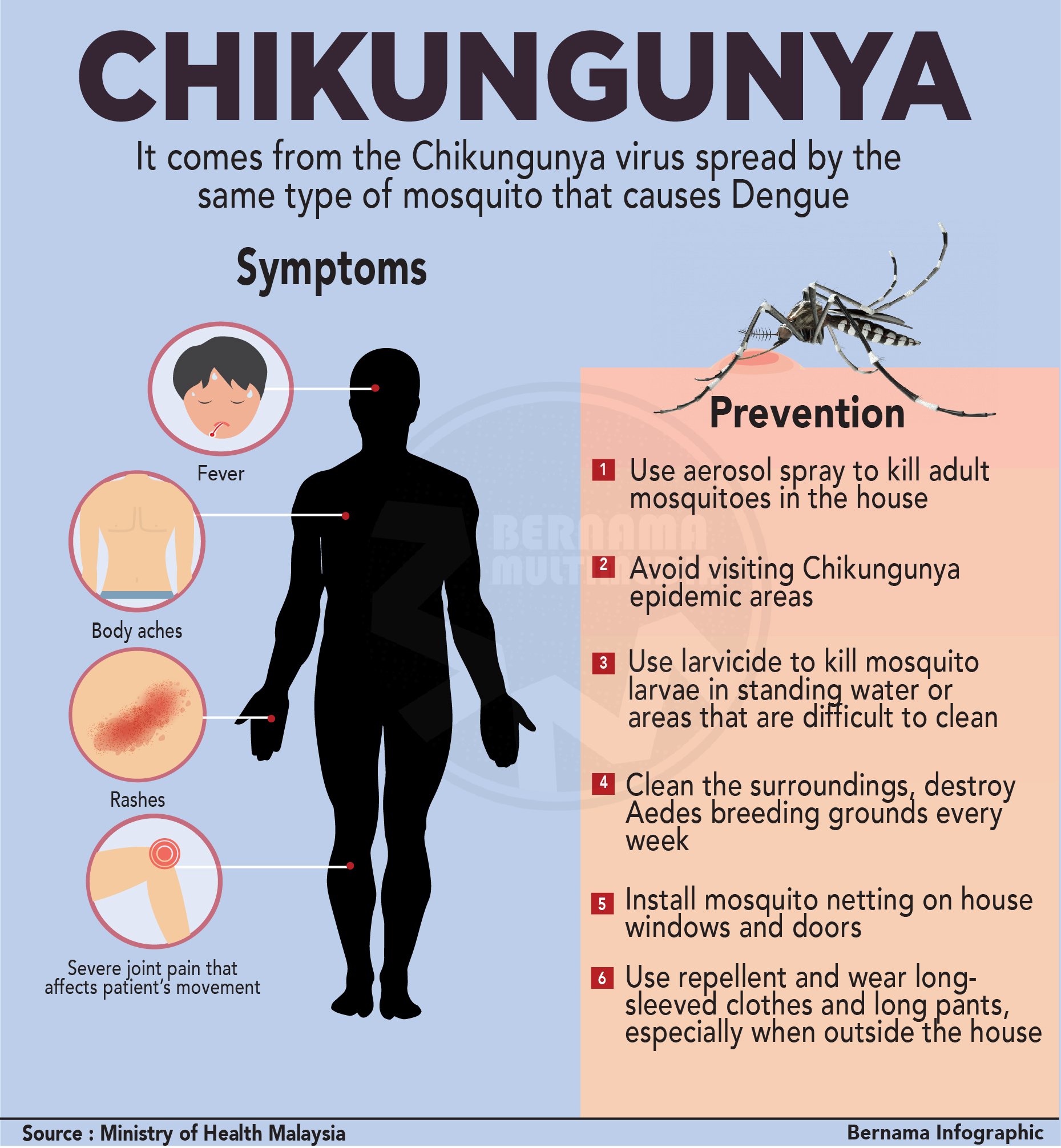
The Chikungunya virus represents a significant global health threat due to its rapid transmission and debilitating clinical manifestations. As an alphavirus within the Togaviridae family, it leverages mosquito vectors such as Aedes aegypti and Aedes albopictus to propagate across tropical and subtropical regions, causing acute febrile illness accompanied by severe arthralgia. Emerging outbreaks in Africa, Asia, and the Americas underscore its adaptive evolutionary trajectory, while diagnostic complexities and limited therapeutic options exacerbate public health burdens. This analysis explores the virus’s virological intricacies, clinical impact, and epidemiological dynamics to inform evidence-based interventions.
Understanding Chikungunya requires examining its molecular architecture, including its single-stranded RNA genome and icosahedral capsid, alongside the pathophysiological mechanisms driving acute and chronic symptoms. Comparative epidemiology reveals overlapping features with dengue and Zika, complicating differential diagnosis and necessitating advanced laboratory techniques. Meanwhile, public health strategies must integrate vector control, surveillance systems, and emerging vaccine candidates to mitigate transmission and reduce long-term morbidity. The interplay between environmental factors, urbanization, and healthcare disparities further shapes regional outbreak patterns, demanding a multidisciplinary approach to containment.
Scientific Overview of the Chikungunya Virus
The Chikungunya virus (CHIKV) represents a significant global health challenge due to its rapid transmission, high morbidity, and expanding geographic range. As an arbovirus primarily transmitted by Aedes mosquitoes, CHIKV exhibits distinct virological and epidemiological characteristics that influence its pathogenicity and spread. This section provides a structured analysis of its taxonomic classification, structural biology, transmission dynamics, and historical outbreak patterns, supported by comparative data to contextualize its public health impact.
Virological Classification and Structural Characteristics
The Chikungunya virus belongs to the Togaviridae family, specifically the Alphavirus genus, which also includes viruses such as Sindbis and Ross River virus. CHIKV is an enveloped, single-stranded positive-sense RNA virus with a genome approximately 11.8 kilobases (kb) in length. Its icosahedral capsid, composed of 240 capsid proteins, encapsulates the genomic RNA, while the envelope is formed by E1 and E2 glycoproteins, critical for receptor binding and membrane fusion during host cell entry.
Key structural features include:
Genome stability and adaptation: CHIKV exhibits high mutation rates in the E1 and E2 genes, enabling rapid adaptation to new vectors (e.g., Aedes albopictus) and contributing to epidemic spread.
Transmission Cycle and Vector Biology
CHIKV transmission is mosquito-borne, with Aedes aegypti and Aedes albopictus serving as primary vectors. The cycle involves:1. Viremic human host: Infected individuals develop viremia (virus in blood) for 2–12 days, peaking during symptom onset.
2. Vector acquisition: Mosquitoes acquire the virus by feeding on viremic hosts, with Ae. albopictus exhibiting higher competence for CHIKV due to its broader geographic distribution.
3. Transstadial transmission: The virus persists across mosquito developmental stages (larvae to adults), ensuring vertical transmission.
4. Human reinfection: Immunity is partial, allowing repeated infections with different strains.
Environmental factors influencing spread:
Zoonotic potential: While primates and birds may act as reservoirs, human-to-mosquito-to-human cycles dominate epidemics, with no evidence of sustained zoonotic maintenance.
Comparative Timeline of Major Outbreaks
CHIKV emerged in 1952–1953 in Tanzania (East Africa) before spreading to Asia and the Pacific. Recent decades have seen escalating epidemics due to globalization and vector adaptation. Below is a chronological summary of key outbreaks:| Year/Period | Region | Notable Features | Epidemiological Impact |
|---|---|---|---|
| 1952–1953 | Tanzania (East Africa) | First documented outbreak; linked to Aedes africanus. | Limited to rural areas; low mortality but high morbidity. |
| 1960s–1970s | Asia (Thailand, India) | Sporadic cases; Ae. aegypti as primary vector. | Endemic in South/Southeast Asia; underreported due to diagnostic challenges. |
| 2004–2006 | Indian Ocean (Réunion) | First large-scale epidemic; E1-A226V mutation enhanced Ae. albopictus transmission. | >250,000 cases; high attack rates (37% in Réunion). |
| 2007–2014 | Europe (Italy, France) | Autochthonous transmission in temperate climates; Ae. albopictus expansion. | >300,000 cases in Italy; first European outbreaks. |
| 2013–2016 | Americas (Caribbean) | Rapid spread via Ae. aegypti; Puerto Rico (2014) saw 28,000 cases. | First major outbreak in the Western Hemisphere; linked to travel and trade. |
| 2017–2023 | Africa (Angola, DR Congo) | Re-emergence with Asian lineage strains; co-circulation of multiple genotypes. | >1 million suspected cases; chronic arthritis in 50% of survivors. |
Key Virological Features of Chikungunya Virus
The following table summarizes critical virological and epidemiological attributes of CHIKV, formatted for responsive display:| Feature | Details | Notes |
|---|---|---|
| Genome Type | Single-stranded, positive-sense RNA | Encodes 12 proteins; nsP3 linked to RNA replication. |
| Genome Size | ~11.8 kb | Largest among alphaviruses; includes 5′ and 3′ untranslated regions (UTRs). |
| Capsid Symmetry | Icosahedral (T=4) | 240 capsid proteins form a protective shell around the RNA. |
| Envelope Proteins | E1 (fusion), E2 (receptor binding), E3 (processing) | E1-A226V mutation enhances Ae. albopictus transmission. |
| Incubation Period | 3–7 days (range: 1–12 days) | Asymptomatic cases may extend detection windows. |
| Viremia Duration | 2–12 days (peaks at symptom onset) | Critical for mosquito infection; declines post-symptoms. |
| Primary Symptoms | Acute: Fever, arthralgia, rash; Chronic: Persistent arthritis (50% of cases) | Arthritis may persist for months/years. |
| Case Fatality Rate | <0.1% (higher in elderly/immunocompromised) | Deaths rare but linked to secondary infections (e.g., sepsis). |
| Serotypes/Genotypes | 3 genotypes (West African, East/Central/South African, Asian) | Asian lineage dominates recent outbreaks; genetic recombination rare. |
| Diagnostic Methods | RT-PCR (acute phase), IgM/IgG ELISA (convalescent phase), viral culture (research) | RT-PCR preferred within 7 days of symptom onset. |
Diagnostic challenge: Cross-reactivity with other alphaviruses (e.g., O’nyong-nyong) complicates serological testing; molecular methods remain gold standard.
Clinical Manifestations and Pathophysiology of Chikungunya Virus Infection
Chikungunya virus (CHIKV) infection presents with a distinct clinical spectrum characterized by acute systemic symptoms, prolonged musculoskeletal complications, and immune-mediated sequelae. The acute phase predominantly features debilitating arthralgia, fever, and rash, driven by viral replication and dysregulated host immune responses. Chronic arthritis/arthralgia in a subset of patients reflects persistent immune activation, joint tissue damage, and potential autoimmune-like mechanisms. Differential diagnosis with dengue and Zika viruses remains critical due to overlapping epidemiological and clinical features, necessitating targeted laboratory confirmation.The pathophysiological underpinnings of CHIKV infection involve direct viral cytopathic effects and indirect immune-mediated damage. Viral non-structural proteins (nsP1–nsP4) play a pivotal role in modulating host immune responses, contributing to disease severity and persistence. Below, the acute-phase symptoms, chronic arthritis mechanisms, and comparative clinical features with dengue and Zika are elaborated, alongside the role of viral proteins in immune evasion.
Acute-Phase Symptoms and Pathophysiological Mechanisms
The acute phase of CHIKV infection typically manifests within 3–7 days of mosquito-borne transmission, with symptoms peaking at 2–5 days post-onset. The triad of high-grade fever, severe arthralgia/arthritis, and maculopapular rash defines the clinical presentation, though myalgia, headache, and conjunctivitis are also common. Fever often exceeds 39°C and may persist for 5–7 days, while arthralgia—particularly affecting small joints (hands, wrists, ankles, and feet)—can be incapacitating, mimicking rheumatoid arthritis.Mechanisms underlying acute symptoms include:
Key distinguishing features of acute CHIKV infection:
Development of Chronic Arthritis/Arthralgia and Immune-Mediated Pathways
Approximately 40–60% of CHIKV-infected individuals develop persistent arthritis or arthralgia lasting weeks to years, with some cases progressing to chronic inflammatory arthritis resembling rheumatoid arthritis. The pathophysiology involves:Risk factors for chronic arthritis:
Diagnostic challenges:
Comparison with Dengue and Zika Viruses: Clinical Overlaps and Differential Diagnosis
CHIKV shares epidemiological and clinical features with dengue virus (DENV) and Zika virus (ZIKV), necessitating laboratory confirmation for accurate diagnosis. Below is a comparative analysis of key clinical and laboratory distinctions:| Feature | Chikungunya Virus | Dengue Virus | Zika Virus |
|---|---|---|---|
| Incubation Period | 3–7 days | 4–10 days | 3–14 days |
| Fever Duration | 5–7 days (often biphasic) | 2–7 days (classic dengue) / prolonged (DHF) | Low-grade or brief |
| Arthralgia | Severe, symmetric, polyarticular (90%+) | Mild to moderate (30–50%) | Mild, transient |
| Rash | Maculopapular, pruritic (70%+) | Maculopapular (50%), petechial (DHF) | Maculopapular, facial flushing |
| Hemorrhagic Manifestations | Rare (uncommon) | Common (DHF/DSS: thrombocytopenia, bleeding) | Rare (mild thrombocytopenia) |
| Neurological Symptoms | Mild (meningitis, encephalitis in severe cases) | Encephalopathy (rare) | Microcephaly (congenital), Guillain-Barré (adults) |
| Chronic Sequelae | Persistent arthritis (40–60%) | Post-dengue fatigue syndrome (PDFS) | Congenital Zika syndrome (microcephaly, ocular defects) |
| Serological Tests | IgM/IgG ELISA (cross-reacts with O’nyong-nyong) | NS1 antigen, IgM/IgG (cross-reacts with DENV) | IgM ELISA (cross-reacts with DENV) |
| PCR Sensitivity | High in acute phase (<7 days) | High in acute phase (<5 days) | High in acute phase (<10 days) |
| Differential Markers | Elevated ferritin, CRP, procalcitonin | Thrombocytopenia, elevated liver enzymes | Elevated ALT, mild leukopenia |
Key distinguishing serological patterns:
Role of Viral Non-Structural Proteins in Immune Evasion and Disease Progression
CHIKV encodes non-structural proteins (nsP1–nsP4), which collectively form the viral replication complex and modulate host immune responses to evade clearance. Their roles in pathogenesis include:The nsP1 protein inhibits type I interferon (IFN-α/β) signaling by degrading STAT2, a critical transcription factor for antiviral responses. nsP2 disrupts mRNA processing and antiviral protein synthesis, while nsP3 interacts with host RNA helicases (e.g., RIG-I) to suppress IFN production. nsP4, the RNA-dependent RNA polymerase, ensures viral replication fidelity but also induces endoplasmic reticulum stress, triggering pro-inflammatory pathways. Collectively, these proteins create an immunosuppressive microenvironment, prolonging viremia and facilitating tissue tropism (e.g., joints, synovium).Mechanisms of immune evasion:
Diagnostic Methods and Laboratory Techniques for Chikungunya Virus
Accurate and timely diagnosis of Chikungunya virus (CHIKV) infection is critical for effective clinical management, epidemiological surveillance, and public health response. The diagnostic approach varies depending on the phase of infection, availability of resources, and clinical presentation. Standard methods include serological assays for antibody detection, molecular techniques for viral RNA identification, and specialized tests for differentiation from other arboviral infections. Each technique has distinct advantages, limitations, and optimal application windows, necessitating a structured diagnostic algorithm tailored to patient presentation and laboratory capabilities.The diagnostic workflow for CHIKV integrates pre-analytical considerations (e.g., sample type, storage, and transport) with analytical precision to minimize false positives or negatives. Molecular assays remain the gold standard during the acute phase, while serological tests are essential for retrospective diagnosis. Advanced techniques, such as next-generation sequencing (NGS) and neutralization assays, provide higher resolution for complex cases or outbreaks involving multiple arboviruses.
Standard Diagnostic Algorithms for Chikungunya Virus
The World Health Organization (WHO) and Centers for Disease Control and Prevention (CDC) recommend a tiered diagnostic approach based on the temporal phase of infection:- Acute Phase (0–5 days post-symptom onset): Viral RNA detection via reverse transcription polymerase chain reaction (RT-PCR) is the preferred method due to high viral load and specificity.
Key Considerations:
Step-by-Step Procedure for Clinical Sample Processing
Proper handling of clinical specimens is essential to ensure diagnostic accuracy and safety. Below is a standardized protocol for processing serum, whole blood, and synovial fluid for CHIKV detection, adhering to biosafety level 2 (BSL-2) guidelines.Pre-Analytical Phase:
Analytical Phase:
Post-Analytical Phase:
Comparison of Diagnostic Methods for Chikungunya Virus
The following table summarizes the key characteristics of diagnostic methods for CHIKV, including sensitivity, specificity, turnaround time, and clinical utility. Data are derived from meta-analyses and WHO/CDC guidelines.| Test Type | Detection Window | Sensitivity (%) | Specificity (%) | Turnaround Time | Cost (USD, approximate) | Clinical Utility | |||||||||||||||||||||
|---|---|---|---|---|---|---|---|---|---|---|---|---|---|---|---|---|---|---|---|---|---|---|---|---|---|---|---|
| RT-PCR (Serum/Whole Blood) | 0–5 days post-onset | 80–95 | 95–100 | 6–24 hours | 20–50 per test |
|
|||||||||||||||||||||
| IgM ELISA (Serum/Plasma) | 6–12 days post-onset | 70–90 | 85–95 | 24–48 hours | 10–30 per test |
|
|||||||||||||||||||||
| IgG ELISA (Serum/Plasma) | ≥12 days post-onset (persists lifelong) | 90–98 | 90–98 | 24–48 hours | 15–40 per test |
|
|||||||||||||||||||||
| PRNT (Serum) | Acute or convalescent phase | 95–100 | 99–100 | 5–7 days | 50–100 per test |
Treatment Strategies and Public Health Interventions for Chikungunya VirusThe Chikungunya virus (CHIKV) infection lacks specific antiviral therapies, necessitating a multifaceted approach combining symptomatic management, experimental interventions, and robust public health measures. While supportive care remains the cornerstone of treatment, ongoing research explores antiviral agents and monoclonal antibodies, while vector control and surveillance systems form the backbone of outbreak mitigation. Vaccination development presents a promising long-term solution, though challenges in strain variability and global deployment persist.Current Therapeutic ApproachesSymptomatic and Supportive ManagementThe primary treatment for CHIKV infection focuses on alleviating clinical manifestations, as no licensed antiviral therapy exists. Nonsteroidal anti-inflammatory drugs (NSAIDs) such as ibuprofen and naproxen are commonly prescribed to reduce fever, arthralgia, and myalgia, though caution is advised in patients with renal impairment or gastrointestinal bleeding risks. Acetaminophen (paracetamol) is preferred in individuals with contraindications to NSAIDs or those with underlying liver disease. Hydration and rest are critical, particularly in severe cases involving dehydration or neurological complications. Physical therapy and assistive devices may be required for prolonged arthritic symptoms, which can persist for months or years in a subset of patients. Experimental and Investigational Treatments Immunomodulatory and Adjuvant Therapies Public Health Strategies for Outbreak MitigationVector Control MeasuresThe primary strategy for preventing CHIKV transmission relies on interrupting the Aedes aegypti and Aedes albopictus mosquito life cycle. Larval habitat reduction is a cost-effective, sustainable approach involving community engagement to eliminate stagnant water sources (e.g., discarded tires, flowerpot saucers, and roof gutters). Insecticide-treated nets (ITNs) and indoor residual spraying (IRS) with pyrethroids or organophosphates reduce human-mosquito contact, though resistance development in Aedes species poses a growing challenge. Biological control methods, such as the release of Wolbachia-infected mosquitoes (e.g., Olyset or Viennot strains), have shown efficacy in suppressing CHIKV transmission in field trials by impairing viral replication in mosquitoes. Environmental management programs (EMPs) integrate GIS mapping, larval surveillance, and targeted interventions in high-risk urban areas. Surveillance and Early Warning Systems Community Engagement and Behavioral Interventions Community-Based Chikungunya Response Plan FlowchartBelow is a text-based pseudocode flowchart outlining a structured community response plan, designed for scalability and adaptability:START Key Features of the Flowchart: Vaccine Development and Deployment ChallengesClinical Trial Phases and Efficacy DataThree CHIKV vaccine candidates have advanced to Phase III trials, with VLA1553 (Valneva) and CVD 1904 (Meiji Seika Pharma) showing the most promising results. VLA1553, a live-attenuated vaccine based on the LR2006-OPY1 strain, demonstrated 100% Epidemiological Trends and Global Impact of Chikungunya VirusThe Chikungunya virus (CHIKV) has emerged as a significant global health threat, reshaping arboviral epidemiology through rapid geographic expansion, shifting transmission dynamics, and disproportionate socioeconomic burdens. Since its initial characterization in 1952, CHIKV has evolved from a localized African and Asian pathogen into a globally circulating virus, facilitated by urbanization, climate variability, and international travel. This section examines the current epidemiological landscape, including incidence patterns, mortality estimates, and socioeconomic consequences, while analyzing emerging trends such as vector adaptation, climate-driven expansion, and the interplay with co-circulating arboviruses. Geographical disparities in healthcare infrastructure further exacerbate the virus’s impact, necessitating a comparative analysis of its effects across income strata.Global Burden and Incidence PatternsChikungunya virus infections have surged since the early 2000s, with the World Health Organization (WHO) reporting over 1.1 million suspected cases between 2005 and 2017, though underreporting likely inflates the true burden. The 2013–2014 epidemic in the Americas marked a turning point, with 1.4 million suspected cases across 46 countries, including 1.3 million in the Americas alone (PAHO, 2014). Incidence rates vary by region, with La Réunion (2005–2006) experiencing an attack rate of 30–40% in some areas, while India’s 2016 outbreak affected 1.5 million individuals in a single year (Ministry of Health and Family Welfare, India, 2016).Mortality from CHIKV remains low (case-fatality rate <0.1% in most outbreaks), but complications—such as severe arthritis, neurological sequelae, and Guillain-Barré syndrome—contribute to long-term morbidity. Disability-adjusted life years (DALYs) attributable to CHIKV are estimated at 1.2 million globally (2000–2016), with 20–30% of infected individuals experiencing persistent symptoms for months to years (Institute for Health Metrics and Evaluation, 2018). Socioeconomic costs include: Emerging Epidemiological TrendsUrbanization and Vector AdaptationThe virus’s spread is closely linked to urbanization, where Aedes aegypti and Aedes albopictus thrive in dense populations with poor sanitation. Key trends include:Climate Change and Vector HabitatsClimate variability directly influences CHIKV transmission by expanding Aedes mosquito ranges. Studies project:Co-Circulation with Other ArbovirusesThe rise of dual or triple arbovirus infections (e.g., CHIKV + dengue + Zika) complicates epidemiology and clinical management. Key observations:Geographical Heatmap: High-Risk Regions and Risk FactorsA text-based global risk stratification reveals distinct transmission hotspots, categorized by urban density, vector presence, and healthcare gaps:
|
Leave a Comment
Comments are moderated before appearing. The data you submit is processed according to the Privacy Policy of Ns2.