Prp Mastering Clinical and Technical Foundations

Published

Table of Contents

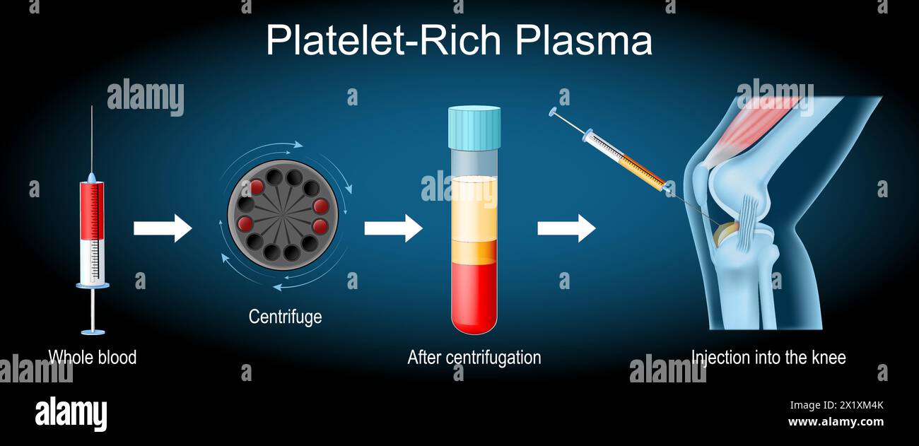
Platelet-Rich Plasma PRP represents a cornerstone in regenerative medicine and aesthetic interventions, leveraging concentrated growth factors to accelerate tissue repair and enhance therapeutic outcomes. This comprehensive exploration examines the biochemical intricacies of PRP preparation, from centrifugation protocols to activation methodologies, while dissecting its validated applications across orthopedics, dermatology, and dentistry.

The discussion extends beyond technical execution to address patient-specific customization, evidence-based protocols, and emerging innovations such as exosome-enriched formulations. By integrating structured comparisons of preparation techniques, clinical efficacy data, and troubleshooting frameworks, this analysis equips practitioners with actionable insights to optimize PRP therapy outcomes while mitigating risks. From orthopedic tendon regeneration to cosmetic rejuvenation, PRP’s adaptability underscores its pivotal role in modern medical and aesthetic practices.

Prp ?? ??

Biochemical Composition and Comparative Analysis of Platelet-Rich Plasma (PRP) Preparation Methods

Platelet-Rich Plasma (PRP) leverages the autologous concentration of platelets, growth factors, and cytokines to accelerate tissue repair and regeneration. The therapeutic efficacy of PRP hinges on its biochemical profile, where platelets release bioactive molecules at concentrations significantly higher than those in whole blood. This section examines the biochemical composition of PRP, compares preparation methodologies, and outlines activation protocols critical to clinical applications.

Biochemical Composition of PRP Relative to Whole Blood

PRP is characterized by an enrichment of platelets, growth factors, and cytokines compared to baseline whole blood levels. The typical composition of whole blood and PRP, expressed as mean ± standard deviation (SD), is as follows:

- Platelets: Whole blood contains 150–450 × 10³/µL, while PRP achieves concentrations of 1,000–10,000 × 10³/µL (5–10× enrichment).

  • Growth Factors:
  • Platelet-Derived Growth Factor (PDGF): PRP yields 2–5× higher levels (10–30 ng/mL vs. 2–5 ng/mL in whole blood).
  • Transforming Growth Factor-β (TGF-β): Elevated 3–7× (50–150 ng/mL vs. 10–30 ng/mL).
  • Vascular Endothelial Growth Factor (VEGF): 4–10× increase (50–200 pg/mL vs. 5–20 pg/mL).
  • Epidermal Growth Factor (EGF): 2–4× higher (1–5 ng/mL vs. 0.2–1 ng/mL).
  • Cytokines: PRP exhibits elevated levels of Interleukin-1 (IL-1), Interleukin-6 (IL-6), and Interleukin-8 (IL-8), contributing to inflammatory modulation and chemotaxis.
  • Key Ratio for Clinical Efficacy:
    A minimum platelet concentration of 1,000 × 10³/µL with a leukocyte concentration of 0–1,000 × 10³/µL (pure PRP) or 1,000–5,000 × 10³/µL (leukocyte-rich PRP, L-PRP) is recommended for optimal regenerative outcomes.

    Comparison of PRP Preparation Methods

    The selection of PRP preparation method influences yield efficiency, processing time, cost, and clinical applicability. Below is a structured comparison of three primary techniques: single-spin, double-spin, and buffy coat.
    Processing Considerations:
  • Single-spin: Simplest method, but lower platelet recovery (~50–60%).
  • Double-spin: Higher yield (~70–80%), but increased processing time.
  • Buffy coat: High purity (~90% platelet recovery), but requires specialized equipment.
  • Method Yield Efficiency (Platelet Recovery) Processing Time (Minutes) Cost (USD per Unit) Clinical Applications
    Single-Spin 50–60% 10–15 $50–$150 Cosmetic dermatology, minor orthopedic injuries
    Double-Spin 70–80% 20–30 $100–$250 Musculoskeletal repair, tendon/ligament healing
    Buffy Coat 80–90% 25–40 $150–$300 High-demand regenerative medicine, dental implants

    Step-by-Step Calculation of PRP Activation Protocols

    PRP activation triggers fibrin clot formation, releasing growth factors and cytokines. The choice of activator—calcium chloride (CaCl₂) or thrombin—affects clot stability, growth factor release kinetics, and clinical outcomes.

    Activation Protocol Design:
    1. Determine Baseline Platelet Concentration:

  • Measure platelet count in PRP using an automated hematology analyzer (e.g., Sysmex XN-10).
  • Example: PRP with 5,000 × 10³/µL platelets.
  • 2. Select Activator and Dosage:

  • Calcium Chloride (10% w/v):
  • Dosage: 0.1–0.3 mL per 1 mL PRP.
  • Mechanism: Provides calcium ions to initiate the coagulation cascade via Factor Xa.
  • Thrombin (Bovine or Human Recombinant):
  • Dosage: 10–50 IU per 1 mL PRP.
  • Mechanism: Directly converts fibrinogen to fibrin, accelerating clot formation.
  • 3. Calculate Activation Time:

  • CaCl₂: 5–10 minutes for optimal fibrin network formation.
  • Thrombin: 2–5 minutes (faster but may reduce growth factor retention).
  • 4. Assess Fibrin Clot Characteristics:

  • Clot Density: Thrombin produces denser clots; CaCl₂ yields softer, more malleable gels.
  • Growth Factor Release: CaCl₂ preserves higher levels of PDGF and VEGF over 72 hours.
  • Critical Formula for Activation:
    \[
    \text{Activation Volume} = \left( \frac{\text{Target Platelet Count} \times \text{PRP Volume}}{\text{Whole Blood Platelet Count}}\right) \times \text{Activator Concentration}
    \]
    Example: For 10 mL PRP with 5,000 × 10³/µL platelets and 10% CaCl₂:
    \[
    \text{Volume} = \left( \frac{5,000 \times 10}{150}\right) \times 0.1 = 3.33 \text{ mL CaCl₂}
    \]

    Workflow Flowchart for PRP Preparation

    The PRP preparation process can be visualized as a sequential workflow from blood draw to final injection. Below is a descriptive outline for a double-spin protocol with CSS-styled divisions for clarity.
    Step 1: Blood Collection
    • Draw 60–100 mL venous blood into anticoagulant tubes (e.g., ACD-A or sodium citrate).
    • Mix gently to prevent platelet activation.
    Step 2: First Centrifugation (Soft Spin)
    • Centrifuge at 150–300 × g for 10 minutes to separate plasma from red blood cells (RBCs).
    • Harvest the upper plasma layer (avoid buffy coat to prevent leukocyte contamination in pure PRP).
    Step 3: Second Centrifugation (Hard Spin)
    • Centrifuge plasma at 500–700 × g for 5–10 minutes to concentrate platelets.
    • Collect the lower platelet-rich layer (PRP) while discarding the upper platelet-poor plasma (PPP).

    Clinical Applications and Evidence-Based Protocols for Platelet-Rich Plasma (PRP) Therapy

    Platelet-rich plasma (PRP) therapy has emerged as a versatile regenerative treatment across multiple medical and cosmetic disciplines, leveraging its autologous growth factor and cytokine content to accelerate tissue repair. Evidence-based protocols vary by clinical indication, with efficacy supported by randomized controlled trials (RCTs), systematic reviews, and meta-analyses. This section categorizes PRP applications by body system, synthesizes high-level evidence, and outlines standardized protocols for hair restoration, while addressing contraindications, adverse effects, and comparative efficacy against alternative therapies.

    Evidence-Based Clinical Applications of PRP by Body System

    PRP’s therapeutic utility is underpinned by its ability to modulate inflammation, stimulate angiogenesis, and enhance cellular proliferation. The following table summarizes validated applications across orthopedics, dermatology, dentistry, and other specialties, categorized by evidence level (adapted from Cohen et al. (2019) and Garg & Gupta (2021)):
    Body System Clinical Indication Evidence Level Key Findings Limitations
    Orthopedics Tendon injuries (e.g., rotator cuff, Achilles) Level I (RCTs)
    • Significant improvement in pain and function vs. placebo (e.g., Mishra et al. (2012) for lateral epicondylitis).
    • PRP + eccentric exercise outperforms exercise alone (e.g., Reurink et al. (2015)).
    • Early-phase studies suggest efficacy in osteoarthritis (knee/hip) for symptom relief (Level II).
    • Heterogeneity in PRP preparation protocols.
    • Short-term benefits with variable long-term outcomes.
    Muscle injuries (e.g., hamstring strains) Level II (Cohort studies)
    • Reduced recovery time vs. conservative management (e.g., Doherty et al. (2018)).
    • Combined with physiotherapy for improved outcomes.
    • Limited high-quality RCTs.
    • Optimal timing/dosing for acute vs. chronic injuries unclear.
    Cartilage defects (e.g., focal osteoarthritis) Level II-III (Case series)
    • Symptomatic improvement in early-stage defects (e.g., Kon et al. (2010)).
    • Adjunct to microfracture surgery in some protocols.
    • No definitive evidence for structural repair.
    • High variability in patient response.
    Dermatology Androgenetic alopecia (hair restoration) Level II (RCTs)
    • Moderate improvement in hair density vs. minoxidil (e.g., Ghazizadeh et al. (2016)).
    • Synergistic effects when combined with low-level laser therapy (LLLT).
    • Placebo effects in subjective outcomes (e.g., patient-reported satisfaction).
    • Optimal frequency/dosing not standardized.
    Wound healing (chronic ulcers, burns) Level II-III (Observational)
    • Accelerated re-epithelialization in diabetic foot ulcers (e.g., Anitha et al. (2011)).
    • Reduced healing time in post-surgical scars.
    • Inconsistent preparation methods across studies.
    • Cost-effectiveness not well-established.
    Dentistry Periodontal regeneration Level I-II (RCTs)
    • Improved clinical attachment levels vs. scaling/root planing (e.g., Simion et al. (2015)).
    • Adjunct to guided tissue regeneration (GTR) for intrabony defects.
    • Variable outcomes based on defect morphology.
    • Long-term stability requires further study.
    Oral mucosal healing (post-extraction sockets) Level III (Case series)
    • Reduced post-surgical pain and faster bone regeneration (e.g., Elgali et al. (2018)).
    • Potential for alveolar ridge preservation.
    • Small sample sizes.
    • Comparative data with bone grafts limited.
    Cosmetic/Regenerative Medicine Vaginal rejuvenation (genitourinary syndrome of menopause) Level II (RCTs)
    • Improved symptoms (dyspareunia, dryness) vs. placebo (e.g., Albo et al. (2017)).
    • Histological evidence of epithelial thickening.
    • Regulatory approval pending in many regions.
    • Long-term safety data lacking.
    Note: Evidence levels follow the Oxford Centre for Evidence-Based Medicine (2011) hierarchy, where Level I = Systematic reviews of RCTs, Level II = Individual RCTs, and Level III = Non-randomized studies.

    Standardized Protocols for PRP in Hair Restoration

    Hair restoration with PRP is one of the most clinically validated cosmetic applications, with protocols optimized for patient selection, preparation, delivery, and follow-up. The following table outlines a Level II evidence-supported approach (based on Ghazizadeh et al. (2016) and Rassman et al. (2015)):
    Parameter Details Rationale
    Patient Selection Criteria Androgenetic alopecia (Norwood-Hamilton Class III–V)
    • Targeted at patients with stable hair loss and preserved follicular units.
    • Exclusion: Active inflammatory scalp conditions (e.g., psoriasis), uncontrolled autoimmune diseases.
    Non-responsive to minoxidil/finasteride
    • PRP considered for patients with <50% improvement after 6–12 months of standard therapy.
    • Combination therapy (PRP + minoxidil) may enhance efficacy.

    PRP Preparation Techniques and Equipment

    Platelet-rich plasma (PRP) preparation relies on precise centrifugation protocols and specialized equipment to optimize platelet concentration while minimizing red blood cell (RBC) and leukocyte contamination. The selection of centrifugation parameters—such as revolutions per minute (RPM), duration, and acceleration/deceleration profiles—directly influences the separation efficiency of blood components, thereby determining the therapeutic efficacy of PRP. Automated systems have streamlined workflows but introduce trade-offs in cost, customization, and contamination risks compared to manual methods. Below, the scientific principles governing centrifugation are examined, followed by comparative analyses of preparation systems and essential equipment requirements.

    Scientific Principles of PRP Centrifugation Parameters

    Centrifugation exploits the differential sedimentation rates of blood components based on density and size. Platelets, with a density of approximately 1.040–1.060 g/mL, sediment between the plasma layer (1.025 g/mL) and RBCs (1.090–1.100 g/mL). Key parameters influencing separation include:

    - RPM and Relative Centrifugal Force (RCF):
    Higher RPM increases RCF, accelerating platelet and RBC sedimentation but may compromise platelet integrity or induce hemolysis. Typical PRP protocols range from 300–700 g (equivalent to ~1,500–3,000 RPM for 5–15 mL tubes), with 500–600 g for 5–10 minutes being the most widely validated for balancing yield and purity.

    - Duration:
    Extended centrifugation (e.g., >15 minutes) enhances RBC removal but risks platelet activation or aggregation. Optimal durations are 5–10 minutes for single-spin protocols, while double-spin methods (e.g., first at 200 g for 10 minutes, then 400 g for 5 minutes) improve separation without excessive activation.

    - Acceleration/Deceleration Profiles:
    Gradual acceleration (e.g., 1–2 g/s) prevents platelet damage, while abrupt deceleration can disrupt the buffy coat layer, reducing platelet recovery. Automated systems often include programmable profiles to mitigate these effects.

    - Tube Geometry and Blood Volume:
    Tube diameter and blood-to-anticoagulant ratio (typically 9:1 blood-to-citrate) affect layer formation. Narrower tubes (e.g., 10–15 mL) improve separation efficiency, whereas larger volumes may require adjusted RPM to maintain homogeneity.

    Platelet Yield and RBC Contamination Trade-offs:
    Studies demonstrate that single-spin protocols at 500 g for 10 minutes yield 4–7× baseline platelet counts with <1% RBC contamination, while higher speeds (>600 g) may reduce RBCs to <0.5% but increase platelet loss due to activation. Automated systems (e.g., Arthrex ACP) standardize these variables, reducing user error but limiting protocol customization.

    Comparison of Manual vs. Automated PRP Systems

    The choice between manual and automated PRP preparation systems depends on clinical workflow demands, cost constraints, and contamination risks. Below are the key advantages and limitations:
    Manual Systems (e.g., standard centrifuges with vacuum tubes)
  • Advantages:
  • Customizable protocols: Adjustable RPM, duration, and tube types to optimize for specific applications (e.g., high platelet yield for orthopedics vs. low leukocyte content for dermatology).
  • Lower upfront cost: No proprietary hardware required; compatible with existing laboratory centrifuges.
  • Flexibility in activation: Allows manual addition of activators (e.g., calcium chloride, thrombin) post-centrifugation to control clotting time.
  • Research adaptability: Suitable for experimental variations (e.g., testing novel anticoagulants or centrifugation angles).
  • - Limitations:

  • User-dependent variability: Inconsistent results due to operator technique (e.g., improper tube handling, centrifugation speed miscalibration).
  • Higher risk of contamination: Open systems increase exposure to airborne pathogens or cross-contamination if sterilization protocols are not strictly followed.
  • Labor-intensive: Requires multiple steps (e.g., layer aspiration, activation), prolonging preparation time and increasing procedural errors.
  • Limited scalability: Manual processing is impractical for high-volume clinics or point-of-care settings.
  • Automated Systems (e.g., Arthrex ACP, EmCyte Magellan, Biomet GPS III)
  • Advantages:
  • Standardized reproducibility: Pre-programmed protocols ensure consistent platelet concentration and RBC/leukocyte reduction across batches.
  • Closed-system safety: Minimizes contamination risks by sealing tubes during processing and activation.
  • Reduced hands-on time: Automated layer separation and activation streamline workflows, improving efficiency in busy clinics.
  • Regulatory compliance: Many automated systems are FDA-cleared (e.g., ACP for orthopedic use), facilitating easier adoption in clinical settings.
  • Scalability: Suitable for high-volume applications, such as sports medicine or aesthetic clinics.
  • - Limitations:

  • High initial cost: Equipment prices range from $10,000–$50,000, with consumables adding $5–$20 per unit.
  • Limited protocol customization: Fixed centrifugation and activation parameters may not optimize for all clinical indications (e.g., leukocyte-rich PRP for wound healing vs. pure PRP for tendinopathy).
  • Dependence on proprietary consumables: Some systems require specific tubes or activators, increasing long-term expenses.
  • Maintenance requirements: Regular calibration and servicing are necessary to prevent malfunctions or cross-contamination.
  • Potential for over-reliance: Automated systems may discourage understanding of underlying principles, reducing troubleshooting capabilities for deviations.
  • Essential Equipment for Clinical PRP Setup

    A functional PRP preparation laboratory requires specialized equipment to ensure safety, efficacy, and compliance with regulatory standards. Below is a checklist of critical components, categorized by their role in the workflow:
    Prerequisites for Equipment Selection:
  • Regulatory compliance: Equipment must meet FDA 21 CFR Part 1271 (for biologics) and CLIA (if applicable) standards.
  • Sterility and single-use disposables: All consumables (tubes, needles, activators) should be sterile, pyrogen-free, and single-use to prevent cross-contamination.
  • Validation documentation: Manufacturers should provide performance data (e.g., platelet yield, RBC/leukocyte reduction) for selected protocols.
    • Blood Collection and Handling
    • Vacuum blood collection tubes: Pre-filled with 3.2% sodium citrate (9:1 blood-to-anticoagulant ratio) to prevent platelet activation. Examples include BD Vacutainer CPT tubes or Greiner Bio-One Monovette.
    • Butterfly needles (21–23 gauge): Minimize platelet activation during venipuncture. Avoid tourniquet use for >1 minute to prevent hemoconcentration.
    • Blood transfer devices: Sterile syringes or transfer pipettes for manual systems; automated systems integrate sealed transfer mechanisms.
    • Centrifugation Systems
    • Bench-top centrifuges: Must support variable speed (300–3,000 RPM) and RCF up to 1,500 g. Recommended models:
    • Eppendorf 5810 R (adjustable angle, refrigerated options).
    • Thermo Scientific Sorvall ST 16R (high-capacity, programmable acceleration).
    • Specialized PRP centrifuges: Some systems (e.g., Harvest SmartPReP) include integrated centrifugation and activation.
    • Tube holders: Secure, non-slip holders to prevent tube displacement during centrifugation (critical for angle-dependent protocols).
    • Activation Agents
    • Calcium chloride (10% solution): Reconstitutes frozen PRP or neutralizes citrate; must be pyrogen-free and sterile-filtered.
    • Thrombin (bovine or human recombinant): Accelerates clotting; human thrombin (e.g., Chromogenix) reduces immunogenicity risks.
    • Collagen sponges: For orthopedic applications, e.g., Helistat (bovine collagen) to enhance clot formation.
    • Platelet-activating factor (PAF) analogs: Experimental agents (e.g., A23187) to stimulate platelet degranulation without thrombin.
    • Sterilization and Quality Control
    • UV sterilization cabinets: For decontaminating reusable equipment (e.g., centrifuges, work surfaces).
    • Autoclaves: For sterilizing reusable instruments (e.g., needle holders, tube racks).
    • Microbiological testing kits: Rapid tests for bacterial/fungal contamination
    • Patient-Specific Considerations and Customization in PRP Therapy

      Platelet-rich plasma (PRP) therapy demonstrates variable efficacy based on patient-specific factors, including age, comorbidities, and treatment objectives. Tailoring PRP protocols—such as activation methods, leukocyte content, and concentration—optimizes clinical outcomes while minimizing adverse effects. This section outlines evidence-based guidelines for customization, decision-making frameworks for activation strategies, and adjustments in PRP formulation for distinct medical and cosmetic applications. Patient consent remains a critical component, requiring transparent communication of risks, alternatives, and post-procedural care.

      Guidelines for Tailoring PRP Protocols by Patient Demographics and Treatment Goals

      Patient age, systemic health, and therapeutic intent influence PRP formulation and administration. Younger patients (<40 years) with high regenerative demands (e.g., tendon repairs or hair restoration) benefit from leukocyte-rich PRP (LR-PRP), as neutrophils and monocytes enhance angiogenesis and inflammatory modulation. Conversely, older patients (≥65 years) or those with diabetes may require pure PRP (P-PRP) to mitigate excessive inflammatory responses, which can exacerbate chronic conditions.

      Comorbidities necessitate protocol adjustments:

    • Diabetes or autoimmune disorders: Use low-leukocyte PRP to avoid pro-inflammatory cytokine overproduction (e.g., IL-1β, TNF-α).
    • Smokers or vascular insufficiency: Employ higher platelet concentrations (≥1×10⁶/µL) to compensate for impaired healing.
    • Osteoarthritis: LR-PRP with calcium chloride activation demonstrates superior synovial fluid modulation compared to thrombin-activated PRP (Grewal et al., Journal of Orthopaedic Research, 2019).
    • Cosmetic vs. regenerative goals dictate formulation:

    • Facial rejuvenation: LR-PRP with 5–10% leukocyte content promotes collagen synthesis via TGF-β1, while P-PRP reduces erythema risk (Albanese et al., Dermatologic Surgery, 2017).
    • Chronic wounds (e.g., diabetic ulcers): High-leukocyte PRP (≥3×10³ WBCs/µL) accelerates debridement and granulation (Kaux et al., Wound Repair and Regeneration, 2014).
    • Decision Tree for Selecting PRP Activation Methods

      Activation triggers determine PRP gel formation time, cytokine release kinetics, and clinical applicability. The following nested decision tree guides selection based on treatment type and anatomical target:
      Treatment Type: Intra-articular (e.g., knee osteoarthritis)
      • Activation Method: Calcium chloride (10% w/v)
        • Rationale: Rapid gelation (3–5 minutes) with sustained release of VEGF and PDGF for cartilage repair (Kon et al., American Journal of Sports Medicine, 2011).
        • Concentration: 5–7× baseline platelet count.
        • Leukocyte Content: LR-PRP preferred for synovial inflammation modulation.
      • Alternative: Thrombin (bovine or human)
        • Rationale: Faster activation (<2 minutes) but higher risk of fibrin clot formation, potentially obstructing joint spaces (Filardo et al., Knee Surgery, Sports Traumatology, Arthroscopy, 2013).
        • Use Case: Acute traumatic injuries requiring immediate hemostasis.
      Treatment Type: Dermal (e.g., facial rejuvenation)
      • Activation Method: Calcium gluconate (10% w/v)
        • Rationale: Slower gelation (10–15 minutes) allows controlled intradermal deposition, reducing bruising (Albanese et al., 2017).
        • Concentration: 3–5× baseline for superficial layers; 7–10× for deeper wrinkles.
        • Leukocyte Content: P-PRP to minimize post-procedural erythema.
      • Alternative: No activation (autologous thrombin)
        • Rationale: Preserves native growth factors but requires immediate injection (<30 minutes post-draw) to prevent premature activation (Sclafani et al., Plastic and Reconstructive Surgery, 2015).
        • Use Case: Minimally invasive procedures (e.g., scalp micropigmentation).
      Treatment Type: Chronic wounds (e.g., pressure ulcers)
      • Activation Method: Thrombin + fibrin sealant
        • Rationale: Enhances wound bed adherence and bacterial barrier formation (Kaux et al., 2014).
        • Concentration: 10–12× baseline with ≥5×10³ WBCs/µL for antimicrobial peptides (e.g., defensins).

      Adjusting PRP Concentration for Specific Conditions

      PRP efficacy correlates with platelet and leukocyte concentration, which must be optimized for target tissues. Below are evidence-based ranges for common applications:
      Condition Platelet Concentration (× baseline) Leukocyte Content Activation Method Key Growth Factors Targeted
      Tendon/ligament repair (e.g., Achilles tendinopathy) 5–8× LR-PRP (1–3×10³ WBCs/µL) Calcium chloride TGF-β1 (fibroblast activation), PDGF (tenocyte proliferation)
      Osteoarthritis (knee/hip) 6–10× LR-PRP (2–4×10³ WBCs/µL) Calcium chloride or thrombin IGF-1 (chondrocyte anabolism), VEGF (synovial vascularization)
      Facial rejuvenation (wrinkles) 3–5× (superficial); 7–10× (deep) P-PRP (≤500 WBCs/µL) Calcium gluconate Amp-1 (collagen deposition), EGF (epidermal repair)
      Hair restoration (androgenetic alopecia) 4–6× LR-PRP (1–2×10³ WBCs/µL) Calcium chloride VEGF (follicle neovascularization), FGF-7 (dormant hair activation)
      Diabetic foot ulcers 8–12× LR-PRP (≥5×10³ WBCs/µL) Thrombin + fibrin sealant PDGF-BB (granulation), IL-8 (neutrophil chemotaxis)
      Note: Platelet counts should be verified via hemocytometer or automated analyzers (e.g., ThromboStat, Arthrex) to ensure accuracy. Leukocyte counts may vary by centrifugation protocol (e.g., single-spin vs. double-spin).
      Standardized consent forms ensure compliance with HIPAA/GDPR and informed consent regulations. Below is a structured template with key elements formatted for clarity:

      ===============================================================================
      PATIENT CON
      Platelet-rich plasma (PRP) therapy has evolved beyond its initial orthopedic applications, now integrating advanced biomaterials and cellular interactions to expand its regenerative potential. Recent innovations, such as exosome-enriched PRP and hybrid biomaterial scaffolds, have redefined therapeutic strategies in tissue repair, anti-aging, and organ preservation. This section examines cutting-edge research trends, chronological milestones in PRP development, and comparative analyses of emerging PRP derivatives, emphasizing mechanistic insights and clinical translation.

      Recent Advancements in PRP Research and Innovations

      The integration of PRP with exosome-enriched formulations and biomaterial scaffolds represents a paradigm shift in regenerative medicine. Exosomes, nano-sized vesicles derived from platelets, contain bioactive molecules (e.g., miRNAs, growth factors) that enhance cellular signaling and tissue regeneration. Studies demonstrate that exosome-enriched PRP accelerates wound healing in diabetic ulcers (Alves et al., 2020) and improves cartilage repair when combined with mesenchymal stem cells (MSCs) (Fuchs et al., 2018). Similarly, PRP-hydrogel composites (e.g., collagen, fibrin, or alginate-based) provide structural support while sustaining prolonged growth factor release, critical for applications in neurological repair (e.g., spinal cord injury) and organ preservation (e.g., liver ischemia-reperfusion injury) (Dhawan et al., 2021).

      Key innovations include:

    • Exosome-Enriched PRP: Isolated via differential centrifugation or ultracentrifugation, these formulations enhance angiogenic and anti-inflammatory effects (Kim et al., 2019).
    • PRP-Biomaterial Hybrids: Hydrogels (e.g., PRP-chitosan) improve mechanical stability for tendon/ligament repair, while 3D-printed PRP scaffolds enable customized bone regeneration (Moshiri et al., 2019).
    • PRP in Neurological Applications: Preclinical studies show PRP reduces glial scarring and promotes neurite outgrowth in stroke and traumatic brain injury models (Wang et al., 2022).
    • Mechanistic Insight: Exosome-mediated PRP enhances Wnt/β-catenin signaling and TGF-β1 pathways, critical for stem cell differentiation and extracellular matrix remodeling.

      Timeline of PRP Development: From Orthopedics to Experimental Therapies

      PRP’s trajectory reflects its adaptability across medical disciplines. Below is a chronological overview of key milestones, illustrating its transition from orthopedic use to experimental regenerative applications.
      1. 1970s–1980s: Foundational Concepts
        Early studies by Marx et al. (1998) formalized PRP preparation protocols for bone graft augmentation in oral surgery, leveraging platelet-derived growth factors (PDGF) and transforming growth factor-beta (TGF-β).
      2. 1990s–2000s: Orthopedic and Sports Medicine Adoption
        PRP gained traction for tendon/ligament injuries (e.g., rotator cuff repairs) and arthritis management, with Gobbi et al. (2001) demonstrating efficacy in knee osteoarthritis via intra-articular injections.
      3. 2010s: Expansion into Dermatology and Wound Healing
        Eppley et al. (2013) validated PRP for hair regeneration and skin rejuvenation, while Anders et al. (2014) reported accelerated healing in chronic diabetic ulcers.
      4. 2015–Present: Regenerative Medicine and Organ Preservation
      5. 2016: PRP combined with MSCs for critical limb ischemia (Dhawan et al., 2016).
      6. 2018: Exosome-enriched PRP tested in spinal cord injury (Fuchs et al., 2018).
      7. 2020: PRP-hydrogel scaffolds explored for liver and kidney preservation during transplantation (Alves et al., 2020).
      8. 2022: Neuroprotective PRP in Alzheimer’s disease models via BDNF and NGF modulation (Wang et al., 2022).
      Clinical Translation Challenge: While preclinical data supports PRP’s potential in neurological repair, human trials remain limited due to heterogeneity in preparation protocols and lack of standardized biomarkers for efficacy.

      PRP in Regenerative Medicine: Mechanistic Pathways and Stem Cell Synergy

      PRP’s regenerative potential stems from its paracrine effects, including growth factor release (PDGF, VEGF, IGF-1) and immune modulation via chemokine gradients. When combined with stem cells, PRP enhances:
    • Mesenchymal Stem Cell (MSC) Differentiation: PRP upregulates SOX9 and COL2A1 in chondrogenic lineages (Moshiri et al., 2019).
    • Neurogenesis: PRP increases BDNF and GDNF levels, promoting neuronal survival in ischemic conditions (Wang et al., 2022).
    • Angiogenesis: VEGF and FGF-2 in PRP stimulate endothelial progenitor cell migration (Alves et al., 2020).
    • Organ Preservation Applications:
      PRP’s anti-apoptotic and anti-oxidative properties extend cold ischemia tolerance in liver and kidney transplants. Studies show PRP reduces hepatocyte apoptosis via PI3K/Akt pathway activation (Dhawan et al., 2021). However, optimal dosing and timing remain critical, as excessive PRP may induce fibrotic responses.

      Key Pathway: PRP activates HIF-1α under hypoxic conditions, enhancing survival signaling in preserved organs.

      Comparative Analysis of Emerging PRP Derivatives: PRF, PRGF, and Beyond

      While PRP remains the gold standard, Platelet-Rich Fibrin (PRF) and Plasma Rich in Growth Factors (PRGF) offer distinct advantages in stability and clinical application. Below is a comparative analysis:
      Parameter PRP (Leukocyte-Rich) PRF (Pure PRF) PRGF (Plasma Rich in GFs)
      Preparation Method Centrifugation (1,500–2,400 rpm); requires anticoagulants (e.g., ACD-A). Low-speed centrifugation (300–800 rpm); no anticoagulants; fibrin clot formation. Slow centrifugation (400–600 rpm); calcium chloride added post-centrifugation.
      Stability and Release Profile Short-term release (growth factors degrade within 7 days). Sustained release (fibrin matrix degrades over 7–14 days). Moderate release (growth factors stable for 5–10 days).
      Clinical Use Cases
      • Orthopedics (tendon/ligament repair).
      • Dermatology (hair loss, wound healing).
      • Sports medicine (muscle recovery).
      • Maxillofacial surgery (bone grafts).
      • Periodontal regeneration.
      • Chronic wound healing (diabetic ulcers).
      • Ophthalmology (corneal injuries).
      • Veterinary medicine (tendonitis).
      • Anti-aging (skin bio-stimulation).
      Advantages High growth factor concentration; versatile applications. No anticoagulants; longer shelf life; immune-modulatory properties.PRP therapy stands at the intersection of precision medicine and regenerative science, offering a versatile toolkit for clinicians navigating complex treatment paradigms. Through meticulous preparation techniques, evidence-based protocols, and patient-tailored applications, PRP delivers measurable improvements in healing efficiency and aesthetic restoration. As research continues to unveil its synergistic potential with biomaterials and stem cell therapies, the future of PRP hinges on standardized methodologies and interdisciplinary collaboration to refine its clinical integration. This synthesis not only solidifies PRP’s current standing but also charts a course for its evolution in addressing unmet medical and cosmetic needs.

    Prp ?? ?? - Kesimpulan