Brain Hemorrhage Shot Types Diagnosis Management
Table of Contents
- Medical Definition and Classification of Brain Hemorrhage
- Anatomical and Physiological Distinctions
- Primary Causes and Age-Related Prevalence
- Comparative Analysis of Clinical Features, Diagnosis, and Prognosis
- Diagnostic Imaging & Radiological Features of Brain Hemorrhage
- Non-Contrast CT Scan Findings in Brain Hemorrhage
- MRI Characteristics of Hemorrhage Over Time
- Advanced Imaging for Vascular Abnormalities
- Interpreting Angiographic Images for Hemorrhage Source Localization
- Emergency Management and Acute Interventions in Brain Hemorrhage
- Initial Emergency Department Stabilization: ABCs and Rapid Assessment
- Surgical Interventions: Indications, Techniques, and Contraindications
- Medical Management: Pharmacological Strategies for Acute Hemorrhage Control
- Complications and Long-Term Sequelae of Brain Hemorrhage
- Mechanisms of Common Complications
- Neurological Deficits by Hemorrhage Location
- Timeline of Complications Post-Hemorrhage
- Rehabilitation & Patient Recovery Protocols in Brain Hemorrhage Survivors
- Multidisciplinary Rehabilitation Framework
- Phase-Specific Rehabilitation Protocols
Brain hemorrhage represents a critical neurological emergency with diverse etiologies and profound clinical implications, demanding precise diagnostic acumen and rapid intervention. This condition encompasses a spectrum of pathologies, from intracerebral bleeding to subarachnoid ruptures, each presenting distinct anatomical challenges and therapeutic considerations. Understanding the underlying mechanisms—ranging from hypertensive vascular damage to traumatic shear forces—is essential for clinicians to implement targeted management strategies that mitigate secondary brain injury and optimize patient outcomes.
The progression of hemorrhage within the cranial cavity triggers a cascade of pathophysiological events, including mass effect, edema formation, and ischemic cascades, which collectively determine prognosis. Advanced imaging modalities, such as non-contrast CT and MRI sequences, serve as indispensable tools in differentiating hemorrhage types, assessing vascular abnormalities, and guiding surgical or endovascular interventions. Meanwhile, emergency protocols must integrate hemodynamic stabilization, anti-hypertensive therapy, and neurocritical care to prevent further neurological deterioration while rehabilitation programs address the long-term functional and psychological sequelae.
Medical Definition and Classification of Brain Hemorrhage
Brain hemorrhage refers to the rupture of blood vessels within or around the brain, leading to acute bleeding that disrupts normal neural function. These hemorrhages are classified based on anatomical location and underlying pathology, each with distinct clinical presentations, diagnostic approaches, and prognostic implications. The primary types—intracerebral hemorrhage (ICH), subarachnoid hemorrhage (SAH), epidural hemorrhage (EDH), and subdural hemorrhage (SDH)—differ in etiology, patient demographics, and secondary injury mechanisms. Understanding these distinctions is critical for timely intervention, as delays in diagnosis or management significantly worsen outcomes, including mortality and long-term disability.
The following sections outline the anatomical and physiological characteristics of each hemorrhage type, their predominant causes across age groups, and a comparative analysis of clinical features, diagnostic modalities, and prognostic factors. Secondary injury mechanisms—such as cerebral edema, mass effect, and ischemic cascades—are also detailed to illustrate the progression of neural damage post-hemorrhage.
Anatomical and Physiological Distinctions
Brain hemorrhages are categorized based on the location of bleeding relative to brain structures and the type of vessel involved (arterial vs. venous). The anatomical distinctions influence symptom presentation, imaging findings, and surgical considerations.- Intracerebral Hemorrhage (ICH):
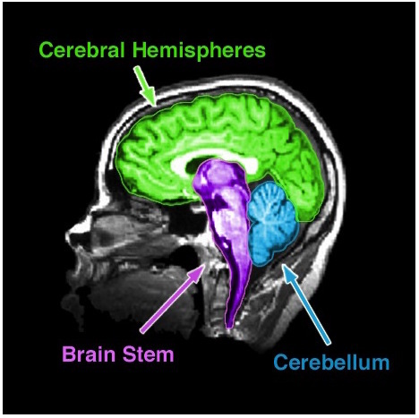
Bleeding occurs within the brain parenchyma, typically originating from small penetrating arteries damaged by hypertension or amyloid angiopathy. The hemorrhage expands into surrounding neural tissue, causing direct compression and edema. Common sites include the basal ganglia, thalamus, cerebellum, and lobes, with each location associated with specific deficits (e.g., cerebellar ICH may cause ataxia or brainstem compression).
- Subarachnoid Hemorrhage (SAH):
Blood accumulates in the subarachnoid space, the cerebrospinal fluid (CSF)-filled area between the arachnoid mater and pia mater. SAH is most frequently caused by ruptured cerebral aneurysms (85% of cases) or arteriovenous malformations (AVMs), though trauma can also contribute. The presence of blood in the CSF triggers vasospasm, hydrocephalus, and chemical meningitis, exacerbating neurological injury.
- Epidural Hemorrhage (EDH):
Blood collects between the dura mater and skull, typically due to trauma (e.g., skull fracture lacerating the middle meningeal artery). The hemorrhage forms a biconvex (lentiform) collection, rapidly increasing intracranial pressure (ICP) as it expands. EDH is less common than subdural hemorrhage but carries a higher risk of herniation if untreated.
- Subdural Hemorrhage (SDH):
Blood accumulates between the dura mater and arachnoid layer, often from bridging veins torn by acceleration-deceleration injuries (e.g., falls, shaken baby syndrome). Chronic SDH may develop insidiously in elderly patients with cerebral atrophy, while acute SDH presents as a crescent-shaped collection on imaging. Venous bleeding results in slower expansion compared to arterial sources.
Primary Causes and Age-Related Prevalence
The etiology of brain hemorrhage varies by type and patient demographics, with hypertension, trauma, and vascular anomalies as the leading contributors. Age-specific patterns emerge due to underlying risk factors:- Intracerebral Hemorrhage (ICH):
- Subarachnoid Hemorrhage (SAH):
- Epidural Hemorrhage (EDH):
- Subdural Hemorrhage (SDH):
Comparative Analysis of Clinical Features, Diagnosis, and Prognosis
The following table summarizes key distinctions between hemorrhage types, including symptoms, diagnostic imaging, and prognostic factors. Understanding these differences guides emergency management and long-term care.| Type of Hemorrhage | Common Symptoms | Imaging Modality | Prognostic Factors | ||||||||||||||||||||||||||||||||
|---|---|---|---|---|---|---|---|---|---|---|---|---|---|---|---|---|---|---|---|---|---|---|---|---|---|---|---|---|---|---|---|---|---|---|---|
| Intracerebral Hemorrhage (ICH) |
|
|
|
||||||||||||||||||||||||||||||||
| Subarachnoid Hemorrhage (SAH) |
|
Endoscopic Evacuation Aneurysm Clipping vs. CoilingContraindications to Surgery Medical Management: Pharmacological Strategies for Acute Hemorrhage ControlMedical therapy complements surgical interventions by controlling blood pressure, reversing anticoagulation, and preventing secondary complications. Targets are tailored to hemorrhage type and patient comorbidities.Blood Pressure Management Mean Arterial Pressure (MAP) Targets by Hemorrhage TypeAntihypertensive Agents and Administration Complications and Long-Term Sequelae of Brain HemorrhageBrain hemorrhage disrupts cerebral homeostasis, leading to a cascade of acute and delayed complications that significantly influence patient prognosis. The pathophysiological consequences vary based on hemorrhage type (e.g., intracerebral, subarachnoid, subdural, epidural), volume, and anatomical location. Early interventions mitigate immediate risks, but delayed sequelae—such as neurocognitive decline, motor deficits, and secondary ischemic events—often determine long-term functional outcomes. Understanding these complications requires integration of neuroanatomical principles, hemodynamic changes, and inflammatory responses.The clinical trajectory post-hemorrhage follows a biphasic pattern: early complications (within 72 hours) arise from mass effect, elevated intracranial pressure (ICP), and systemic instability, while delayed sequelae (days to years) emerge due to cerebral edema, vasospasm, and structural reorganization. Below, the mechanisms, anatomical correlations, and prognostic frameworks are systematically outlined. Mechanisms of Common ComplicationsComplications in brain hemorrhage stem from primary injury (direct tissue destruction) and secondary cascades (e.g., excitotoxicity, oxidative stress, blood-brain barrier disruption). The following pathways underlie the most clinically significant sequelae:- Hydrocephalus - Delayed Cerebral Ischemia (DCI) and Vasospasm - Epileptogenesis - Cognitive Decline and Dementia Neurological Deficits by Hemorrhage LocationThe functional impact of brain hemorrhage correlates with anatomical disruption of white matter tracts, nuclei, or cortical regions. Below is a categorized summary of deficits based on hemorrhage localization:Timeline of Complications Post-HemorrhageThe progression of complications follows a non-linear, multifactorial trajectory influenced by hemorrhage type and patient comorbidities. Below is a stylized flowchart (descriptive text) outlining the critical windows and interventions:
0–72 Hours: Acute Phase Rehabilitation & Patient Recovery Protocols in Brain Hemorrhage SurvivorsThe recovery trajectory following a brain hemorrhage is highly individualized, requiring a multidisciplinary rehabilitation approach to address physical, cognitive, and psychosocial deficits. Survivors often experience motor impairments, cognitive decline, communication difficulties, and emotional disturbances, necessitating tailored interventions across acute, subacute, and chronic phases. Evidence-based protocols integrate neurological recovery principles, functional restoration, and patient-centered adaptation to optimize long-term outcomes. The effectiveness of rehabilitation depends on early initiation, hemorrhage-specific considerations (e.g., subarachnoid vs. intracerebral), and continuous reassessment of patient-specific factors such as age, premorbid function, and comorbidities.Rehabilitation programs must align with neuroplasticity principles, where repetitive, task-specific training enhances cortical reorganization. For example, survivors of intracerebral hemorrhage (ICH) may require aggressive motor retraining due to focal deficits, while subarachnoid hemorrhage (SAH) survivors may prioritize cognitive and vestibular rehabilitation secondary to global cerebral hypoperfusion. Assistive technologies and environmental modifications further bridge the gap between residual deficits and functional independence. Multidisciplinary Rehabilitation FrameworkA structured rehabilitation program integrates physical therapy (PT), occupational therapy (OT), speech-language pathology (SLP), neuropsychology, and social work to address the holistic needs of survivors. Each discipline contributes specialized interventions targeting distinct domains:- Physical Therapy (PT) focuses on motor recovery, balance, and gait training, particularly for survivors with hemiparesis or ataxia. The following table outlines phase-specific therapeutic goals, assistive devices, and recovery timelines, stratified by hemorrhage type and patient factors. Phase-Specific Rehabilitation Protocols
|


Leave a Comment
Comments are moderated before appearing. The data you submit is processed according to the Privacy Policy of Ns2.