Measles Virus Classification Structure Transmission Dynamics
Table of Contents
- Scientific Overview of the Measles Virus
- Taxonomic Classification and Genetic Structure
- Historical Milestones in Measles Virus Research
- Comparative Analysis of Measles Virus with Other Paramyxoviruses
- Intracellular Life Cycle and Immune Evasion
- Transmission, Epidemiology, and Outbreak Dynamics of Measles Virus
- Primary Modes of Measles Transmission
- Factors Influencing Measles Outbreak Dynamics
- Incubation Period and Clinical Progression: Flowchart Analysis
- Clinical Manifestations and Complications of Measles Virus
- Progression of Measles Symptoms: From Infection to Recovery
- Severe Complications and Pathogenesis
- Comparison of Measles Symptoms in Children vs. Adults
- Diagnostic Criteria for Measles
- Diagnostic Tools and Laboratory Techniques for Measles Virus Detection
- Workflow for Measles Diagnosis in Clinical Settings
- Comparison of Diagnostic Methods: Sensitivity, Specificity, and Applicability
- Case Studies: Misdiagnosis and Its Impact on Public Health Responses
The measles virus remains one of the most contagious pathogens globally, with a single infection capable of generating 12 to 18 secondary cases in unvaccinated populations. As a member of the Morbillivirus genus within the Paramyxoviridae family, its single-stranded RNA genome encodes critical proteins—such as hemagglutinin and fusion—that facilitate host cell invasion and immune evasion. From its historical identification in the 19th century to modern genomic sequencing, advancements in virology have illuminated its complex life cycle, transmission pathways, and devastating epidemiological impact.
This analysis explores the virus’s taxonomic and structural intricacies, tracing its evolutionary milestones alongside comparative insights into paramyxovirus relatives. It examines outbreak dynamics shaped by vaccination gaps, population density, and seasonal patterns, while dissecting clinical trajectories from prodromal symptoms to severe complications like encephalitis. Diagnostic challenges, from PCR-based detection to rapid antigen tests, are evaluated within the context of resource-limited settings, underscoring the need for adaptive public health strategies.

Scientific Overview of the Measles Virus
The measles virus (Measles morbillivirus) is a highly contagious pathogen belonging to the Paramyxoviridae family, posing significant global health challenges due to its high transmission efficiency and severe clinical manifestations. Its taxonomic classification, genetic architecture, and evolutionary history reflect its role as a model for understanding paramyxovirus biology. This section explores its phylogenetic placement, genetic organization, historical milestones in its study, and comparative structural features within the Paramyxoviridae family, alongside its intracellular life cycle and immune evasion strategies.Taxonomic Classification and Genetic Structure
The measles virus is classified under the following taxonomic hierarchy:Its genome consists of a non-segmented, single-stranded, negative-sense RNA (~15,900 nucleotides) encoding six structural proteins:
Key Feature: The measles virus exhibits genomic plasticity, with occasional recombination events observed between closely related morbilliviruses (e.g., canine distemper virus).
Historical Milestones in Measles Virus Research
The study of measles spans centuries, from clinical descriptions to molecular characterization. Key developments include:- Ancient Observations (Pre-10th Century): Early records of measles-like illnesses appear in Chinese medical texts (Huangdi Neijing, ~3rd century BCE) and Hippocratic writings (~5th century BCE), though differentiation from rubella and smallpox was unclear.
Significant Impact: The measles vaccine remains one of the most effective public health interventions, with the WHO’s Measles and Rubella Initiative achieving >85% global coverage in high-income countries.
Comparative Analysis of Measles Virus with Other Paramyxoviruses
The Paramyxoviridae family includes pathogens with diverse host ranges and transmission methods. Below is a comparative table highlighting structural, epidemiological, and host-specific differences:| Feature | Measles Virus | Mumps Virus | Respiratory Syncytial Virus (RSV) | Canine Distemper Virus (CDV) |
|---|---|---|---|---|
| Genus | Morbillivirus | Rubulavirus | Pneumovirus | Morbillivirus |
| Genome Size (nt) | 15,900 | 15,384 | 15,222 | 15,690 |
| Surface Glycoproteins | H (hemagglutinin), F (fusion) | HN (hemagglutinin-neuraminidase), F | G (attachment), F | H, F |
| Primary Receptor | SLAM (CD150), nectin-4 | CD46, sialic acid | ICAM-1, heparin sulfate | SLAM, nectin-4 (similar to measles) |
| Transmission Route | Aerosol droplets (highly contagious, R₀ ~12–18) | Saliva/respiratory droplets (R₀ ~4–7) | Direct contact, respiratory droplets (R₀ ~2–5) | Respiratory secretions, direct contact (canine) |
| Host Range | Humans (narrow) | Humans, some primates | Humans, cattle (bovine RSV) | Canids, primates (zoonotic potential) |
| Pathogenesis Focus | Immunosuppression, giant cell formation (syncytia) | Orchitis, meningitis | Bronchiolitis, pneumonia (infants) | Neurological disease, dermatological lesions |
| Vaccine Availability | Live-attenuated (MMR vaccine) | Live-attenuated (Jeryl Lynn strain) | None (preventive monoclonal antibodies in development) | Live-attenuated (canine distemper vaccine) |
Note: While measles and CDV share the same genus and receptors, CDV exhibits broader host adaptation, including zoonotic spillover (e.g., from dogs to big cats in captivity).
Intracellular Life Cycle and Immune Evasion
The measles virus employs a multi-step replication cycle optimized for persistence and immune evasion. Key phases include:1. Attachment and Entry
2. Uncoating and Transcription
3. Replication and Assembly

Transmission, Epidemiology, and Outbreak Dynamics of Measles Virus
The measles virus exhibits high transmissibility due to its prolonged airborne persistence, efficient droplet nucleation, and susceptibility of unvaccinated populations. Understanding its epidemiological behavior—including transmission pathways, incubation phases, and outbreak triggers—is critical for public health interventions. This section examines the primary modes of viral spread, the factors driving outbreaks, and the mathematical frameworks used to model measles dynamics, with comparisons to historical and recent epidemics.Primary Modes of Measles Transmission
Measles spreads primarily through direct contact with infectious respiratory droplets and airborne transmission, with secondary routes involving fomite contamination and environmental persistence. The virus’s stability in aerosols and on surfaces varies by conditions, influencing outbreak risk in confined or densely populated settings."Measles is among the most contagious human pathogens, with an R₀ (basic reproduction number) of 12–18 in unvaccinated populations."Transmission Mechanisms:
— World Health Organization (WHO) and Centers for Disease Control and Prevention (CDC)
- Direct Contact with Secretions
- Fomite Contamination (Indirect Transmission)
"Secondary attack rates in households exceed 90% among susceptible contacts, underscoring the virus’s efficiency in closed settings."
— Study: Journal of Infectious Diseases (2015)
Factors Influencing Measles Outbreak Dynamics
Outbreaks emerge when vaccination coverage drops below herd immunity thresholds, combined with population density, mobility, and seasonal factors. Comparative analysis of historical (pre-vaccine era) and recent outbreaks (e.g., Samoa 2019, Philippines 2023) reveals consistent patterns despite advances in healthcare.Key Drivers of Outbreaks:
-
Vaccination Coverage and Herd Immunity
- Threshold for Elimination: Measles requires ≥95% vaccination coverage to interrupt transmission (herd immunity threshold).
- Critical Gaps:
- Vaccine Hesitancy: Declines in MMR (measles-mumps-rubella) uptake due to misinformation (e.g., 2019 Samoa outbreak linked to anti-vaccine campaigns; coverage dropped from 88% to 31%).
- Missed Opportunities: Low routine immunization in low-income countries (e.g., Nigeria, DRC) fuels regional outbreaks.
- Data: 2023 Philippines saw 10,000+ cases with 70% coverage in some regions, highlighting vulnerability in high-density urban slums.
-
Population Density and Mobility
- Urban Slums: Crowded housing (e.g., Manila, Dhaka) accelerates transmission via prolonged household exposure.
- Travel-Related Spread: International travel reintroduces measles into eliminated regions (e.g., 2019 New Zealand outbreak from travel-related importation).
- Refugee Camps: Displaced populations with low vaccination (e.g., Syrian refugee camps) experience R₀ > 20 due to forced proximity.
-
Seasonal and Climatic Variations
- Winter/Spring Peaks: Outbreaks surge in temperate climates (e.g., U.S. winter 2019) due to indoor crowding and lower UV inactivation of airborne virus.
- Tropical Regions: Year-round transmission in equatorial zones (e.g., Amazon basin) with bimodal peaks linked to rainy seasons (increased indoor gatherings).
- Humidity Impact: Low humidity (<40%) enhances aerosol stability, prolonging infectiousness in dry climates.
-
Healthcare System Resilience
- Diagnostic Delays: Underreporting in resource-limited settings (e.g., India’s 2018 outbreak) due to misdiagnosis as dengue or chickenpox.
- Surge Capacity: Hospitals in low-income countries face overwhelming ICU demand during outbreaks (e.g., Samoa 2019: 83 deaths, 5% case-fatality rate).
| Outbreak | Year | Cases | Vaccination Coverage | Key Trigger | Outcome |
|---|---|---|---|---|---|
| Samoa | 2019 | <17,800 | 31% (MMR1) | Anti-vaccine movement, misinformation | 83 deaths (5% CFR), emergency mass vaccination |
| Philippines (NCR) | 2023 | >10,000 | 70% (regional variation) | Low routine immunization, urban density | 300+ deaths, school closures, supplemental campaigns |
| Madagascar | 2018–2019 | 118,000 | 60% (MMR1) | Displaced populations, weak healthcare | 1,700 deaths, WHO emergency response |
Incubation Period and Clinical Progression: Flowchart Analysis
The measles virus follows a predictable incubation and symptomatic trajectory, with distinct phases marked by viral replication, immune response, and rash onset. Below is a staged flowchart with clinical annotations:[Measles Virus Transmission → Incubation Period (7–21 days)]
│
├── Viral Replication Phase (Days 0–10)
│ ├── Infection Site: Nasopharyngeal mucosa → viremia (Day 7–10).
│ ├── Symptoms: Asymptomatic; viral load peaks in respiratory secretions.
│ └── Infectiousness: Not yet contagious (prodrome begins post-viremia).
│
├── Prodromal Phase (Days 10–14)
│ ├── Day 10–12: Fever (38–40°C), coryza (runny nose), conjunctivitis.
│ ├── Day 12–14: Koplik spots (pathognomonic):
│ │ ├── Location: Buccal mucosa (opposite molars).
│ │ ├── Appearance: 2–3 mm white papules with erythematous base.
│ │ └── Duration: 12
Clinical Manifestations and Complications of Measles Virus
The measles virus (Measles morbillivirus) induces a highly contagious systemic infection characterized by a progressive and distinct clinical trajectory, ranging from mild prodromal symptoms to severe, life-threatening complications. Clinical manifestations are categorized into systemic (e.g., fever, respiratory symptoms) and dermatological (e.g., maculopapular rash) phases, with variations in immunocompromised hosts due to impaired immune responses. Severe complications—such as encephalitis, pneumonia, and subacute sclerosing panencephalitis (SSPE)—arise from viral pathogenesis, immune-mediated damage, or persistent infection. Diagnostic differentiation relies on clinical patterns, laboratory confirmation (e.g., PCR, IgM serology), and exclusion of mimicking exanthematous diseases.Progression of Measles Symptoms: From Infection to Recovery
Measles follows a three-phase clinical course: the incubation period (7–21 days post-exposure), the prodromal phase (2–4 days of systemic symptoms), and the exanthematous phase (rash development). Immunocompromised individuals may exhibit atypical or prolonged presentations, including modified rash patterns, absence of Koplik’s spots, or persistent viremia without overt symptoms.Key Timeframes:Systemic Symptoms (Prodromal Phase):
Incubation: 7–14 days (virus replicates in respiratory mucosa). Prodrome: 2–4 days (viremia peaks; symptoms appear). Exanthem: 3–5 days (rash onset marks infectious peak).
The prodrome begins with high-grade fever (38.3–40.5°C), often accompanied by:
Dermatological Manifestations (Exanthematous Phase):
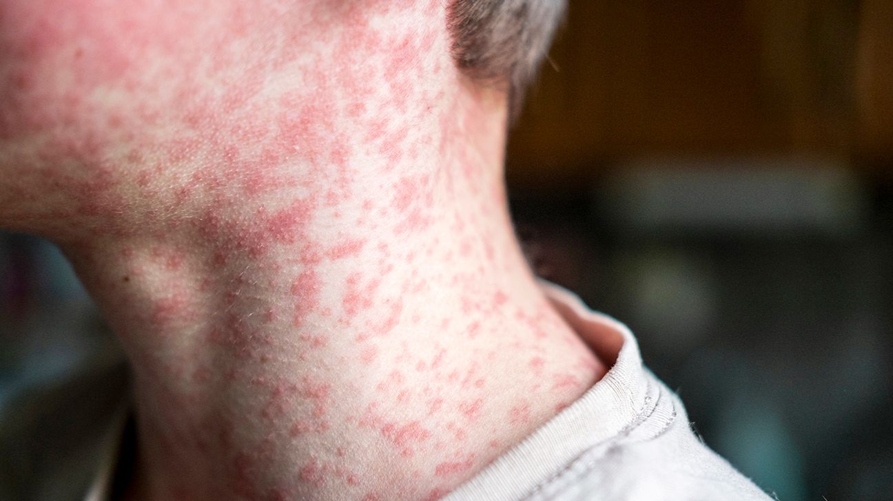
The maculopapular rash emerges 3–5 days post-prodrome, starting at the hairline and spreading centripetally (face → trunk → extremities). Rash characteristics include:
Atypical Presentations in Immunocompromised Hosts:
Severe Complications and Pathogenesis
Measles complications arise from direct viral cytopathology, immune-mediated damage, or persistent infection. The most critical include acute encephalitis, pneumonia, and subacute sclerosing panencephalitis (SSPE), each with distinct mechanisms and sequelae.1. Acute Measles Encephalitis (AME):
2. Measles-Associated Pneumonia:
3. Subacute Sclerosing Panencephalitis (SSPE):
Comparison of Measles Symptoms in Children vs. Adults
Adults experience more severe and frequent complications than children due to waning immunity, higher rates of comorbidities, and delayed diagnosis. The following table summarizes key differences:| Feature | Children (1–14 years) | Adults (≥15 years) |
|---|---|---|
| Prodromal Symptoms | Fever, cough, coryza, conjunctivitis; Koplik’s spots in ~70%. | More severe symptoms (e.g., high fever, diarrhea, dehydration); Koplik’s spots in <50%. |
| Rash Duration | 5–6 days; less confluent. | Longer duration (7+ days); often more confluent/hemorrhagic. |
| Complication Frequency |
|
|
| Atypical Presentations | Rare; modified rash in malnourished. | Frequent (e.g., rash without prodrome, giant-cell pneumonia). |
| Post-Infection Sequelae | Mild (e.g., vitamin A deficiency exacerbation). | Severe (e.g., SSPE, chronic respiratory compromise). |
Diagnostic Criteria for Measles
Measles diagnosis combines clinical suspicion, epidemiological links, and laboratory confirmation. Differentiation from other exanthematous diseases (e.g., rubella, scarlet fever) relies on rash morphology, prodromal symptoms, and laboratory tests.Clinical Diagnosis (WHO Case Definition):
Laboratory Tests:
-
Viral Detection:
- RT-PCR (nasopharyngeal/oral swabs, urine, or blood; gold standard in early infection).
- Viral culture (slow; used for research).
-
Serology:
- IgM antibodies (appears 3–5 days post-rash; persists 1–3 months).
- IgG antibodies (rises post-IgM; lifelong immunity). -
- Use of sterile, single-use collection devices to prevent cross-contamination.
- Immediate placement of specimens in viral transport media (VTM) for throat swabs or urine, with refrigeration or freezing as needed.
- Labeling with patient identifiers, date, and time of collection, along with clinical details (e.g., rash onset, vaccination history).
- Transport in sealed, leak-proof containers with biohazard markings, accompanied by a chain-of-custody form documenting handlers and transfer points.
- Documentation: A signed chain-of-custody form tracks the specimen from collection to disposal, including the names of all handlers, dates, and times of transfer.
- Secure Storage: Specimens are stored in locked refrigerators or freezers with restricted access, with backup power systems for temperature-sensitive samples.
- Transport Regulations: Compliance with international air transport regulations (e.g., IATA) for shipping infectious substances, including proper packaging (e.g., Category B for measles specimens).
- Disposal: Inactivation of specimens (e.g., via chemical treatment or autoclaving) before disposal, with records maintained for audits.
- Cost: RADTs and ELISA kits are more affordable than RT-PCR or virus isolation, though repeated testing may offset initial savings.
- Infrastructure: RT-PCR requires electricity, refrigeration, and trained technicians, while RADTs can be performed in field clinics.
- Training: Serological assays demand technical expertise, whereas RADTs can be deployed by non-laboratory staff after minimal training.
- Cold Chain: Many assays require specimen storage at -20°C or -80°C, which may be unavailable in remote areas.
- Rapid Antigen Tests: High specificity but lower sensitivity, leading to false negatives in early or late infection stages. False positives are rare but can occur due to cross-reactivity with other paramyxoviruses.
- RT-PCR: Highly sensitive and specific but impractical for large-scale screening due to cost and infrastructure needs. False negatives may arise from improper specimen handling or low viral loads.
- Serology: Useful for confirming past infection but limited by the window of IgM detection and potential cross-reactivity with rubella or parvovirus B19.
- Overlap with endemic diseases: Measles symptoms were dismissed due to the presence of dengue and malaria in the region.
- Limited laboratory capacity: Only a small fraction of suspected cases were tested, relying instead on clinical judgment.
- Vaccine hesitancy: A resurgence in anti-vaccination sentiment contributed to low immunization coverage, exacerbating the outbreak. The outbreak prompted a national emergency response, including mass vaccination drives and the deployment of rapid antigen tests to frontline clinics. Post-mortem analyses revealed that 90% of deaths could have been prevented with timely measles-specific interventions.
The measles virus exemplifies the intersection of virology, epidemiology, and global health, where scientific precision and proactive measures determine outbreak control. Its resilience in under-vaccinated communities highlights the fragility of herd immunity thresholds, while diagnostic innovations offer hope for early intervention. As vaccination campaigns and surveillance systems evolve, understanding this pathogen’s mechanisms—from molecular structure to clinical manifestations—remains critical in mitigating its enduring threat. The lessons drawn here reinforce the necessity of sustained research, equitable access to diagnostics, and coordinated public health responses to safeguard vulnerable populations.
Diagnostic Tools and Laboratory Techniques for Measles Virus Detection
Accurate and timely diagnosis of measles remains critical for controlling outbreaks, preventing complications, and guiding public health interventions. The diagnostic workflow for measles integrates clinical suspicion, specimen collection, laboratory processing, and confirmatory testing, with variations in sensitivity, specificity, and feasibility across different settings. Resource constraints in low-income regions often necessitate the use of rapid, cost-effective assays, while high-resource laboratories may employ highly sensitive molecular techniques. Diagnostic errors, particularly misdiagnosis due to overlapping symptoms with other viral illnesses, have historically contributed to delayed responses and prolonged transmission chains.The selection of diagnostic tools depends on the stage of infection, specimen availability, and laboratory infrastructure. Early detection relies on viral RNA or antigen presence, while serological assays confirm exposure or immunity. Emerging technologies, such as CRISPR-based diagnostics and next-generation antigen tests, are refining measles detection, though their implementation in resource-limited environments faces challenges related to cost, training, and cold chain requirements.
Workflow for Measles Diagnosis in Clinical Settings
The diagnostic process for measles begins with clinical assessment and specimen collection, followed by laboratory analysis and interpretation. Proper adherence to chain-of-custody protocols is essential for maintaining sample integrity, particularly when handling infectious materials. The workflow is structured to balance speed, accuracy, and safety, with adjustments made based on the epidemiological context (e.g., outbreak vs. sporadic cases).Specimen Collection and Handling
Specimens for measles diagnosis include throat swabs, urine, and blood, with selection guided by the phase of infection and available resources. Throat swabs are preferred during the acute phase (3 days before to 4 days after rash onset) for viral RNA or antigen detection, while urine specimens may be used for prolonged viral shedding in immunocompromised individuals. Blood samples are collected for serological testing, including IgM and IgG antibody detection. Chain-of-custody protocols must be strictly followed, including:
Laboratory Processing and Testing
Laboratory confirmation of measles involves a tiered approach, combining rapid tests for initial screening and confirmatory assays for definitive diagnosis. The workflow typically includes:
1. Rapid Antigen Detection Tests (RADT): Used in point-of-care settings for preliminary screening, with results available within 30–60 minutes. These tests detect the measles virus nucleocapsid protein in throat swabs or urine, though sensitivity decreases after rash onset.
2. Reverse Transcription Polymerase Chain Reaction (RT-PCR): The gold standard for viral RNA detection, offering high sensitivity and specificity. RT-PCR is performed on throat swabs or urine, with real-time assays enabling quantification of viral load. Limitations include higher costs and the need for specialized equipment.
3. Serological Assays: Enzyme-linked immunosorbent assays (ELISA) or immunofluorescence assays (IFA) detect measles-specific IgM and IgG antibodies. IgM appears 3–5 days after rash onset and persists for 1–3 months, while IgG indicates past infection or vaccination. Serology is less useful in the early phase but confirms exposure in suspected cases with negative viral tests.
Chain-of-Custody for Infectious Samples
Maintaining an unbroken chain of custody is critical for legal, epidemiological, and safety reasons. Protocols include:
Comparison of Diagnostic Methods: Sensitivity, Specificity, and Applicability
The choice of diagnostic method for measles depends on the phase of infection, available resources, and the need for rapid results. Each technique has distinct advantages and limitations, particularly in resource-limited settings where infrastructure and training may be constrained.Performance Characteristics of Diagnostic Assays
The following table compares key diagnostic methods for measles, highlighting their sensitivity, specificity, turnaround time, and suitability for different settings:
| Method | Sensitivity | Specificity | Turnaround Time | Resource Requirements | Best Use Case |
|---|---|---|---|---|---|
| Rapid Antigen Test (RADT) | 50–75% (varies by brand) | 95–99% | 30–60 minutes | Minimal (point-of-care) | Early screening in outbreaks, low-resource settings |
| RT-PCR | 95–100% | 99–100% | 4–24 hours | High (specialized lab, equipment, trained staff) | Confirmatory diagnosis, research, early detection |
| IgM ELISA | 85–95% | 95–98% | 24–48 hours | Moderate (basic lab, reagents) | Retrospective diagnosis, seroprevalence studies |
| IgG ELISA | 90–98% | 98–100% | 24–48 hours | Moderate (basic lab, reagents) | Immunity assessment, vaccination verification |
| Virus Isolation | 90–95% | 100% | 7–14 days | Very high (BSL-3 lab, cell culture) | Rarely used; reference laboratories only |
In low-resource environments, the feasibility of measles diagnosis is often dictated by:
Trade-offs in Diagnostic Accuracy
Case Studies: Misdiagnosis and Its Impact on Public Health Responses
Misdiagnosis of measles, often due to overlapping clinical features with other febrile illnesses (e.g., rubella, dengue, or scarlet fever), has led to delayed interventions and prolonged outbreaks. Below are case studies illustrating the consequences of diagnostic errors and their role in shaping public health responses.Case Study 1: The 2019 Samoa Measles Outbreak
In late 2019, Samoa experienced one of the deadliest measles outbreaks in decades, with 72 deaths among children under 5. Initial misdiagnosis of measles as dengue or chickenpox delayed vaccination campaigns and quarantine measures. Health authorities attributed the diagnostic errors to:
Case Study 2: The 2017 New York City Outbreak
During a measles resurgence in Orthodox Jewish communities in New York, misdiagnosis of measles as "hand, foot, and mouth disease" (HFMD) led to underreporting and delayed contact tracing. Key factors included:
Leave a Comment
Comments are moderated before appearing. The data you submit is processed according to the Privacy Policy of Ns2.