Cervical Adenopathies Diagnosis And Management From Pathophysiology To Th
Table of Contents
- Clinical Definition and Pathophysiology of Cervical Adenopathies
- Anatomical Distribution and Drainage Territories of Cervical Lymph Nodes
- Pathophysiological Mechanisms of Lymph Node Enlargement
- Differentiating Reactive Hyperplasia from Pathological Adenopathy
- Comparative Table: Types of Cervical Adenopathy
- Etiological Classification and Differential Diagnosis of Cervical Adenopathies
- Infectious Etiologies Stratified by Age and Geographic Prevalence
- Neoplastic Etiologies and Risk Factors
- Diagnostic Workflow: From History to Advanced Imaging in Cervical Adenopathy
- Physical Examination: Palpation Techniques and Node Characteristics
- Role of Ultrasound in Cervical Adenopathy Evaluation
- Computed Tomography (CT) and Magnetic Resonance Imaging (MRI) in Cervical Adenopathy
- Red Flags Requiring Immediate Referral
- Therapeutic Strategies by Etiology in Cervical Adenopathies
- Bacterial Cervical Adenitis: Etiology-Specific Antibiotic Protocols
- Viral Cervical Adenopathy: Supportive Care and Antiviral Indications
- Neoplastic Cervical Adenopathy: Staging and Treatment Algorithms
- Comprehensive Treatment Decision Table
- Complications and Prognostic Indicators in Cervical Adenopathies
- Anatomical Complications of Untreated Cervical Adenopathy
- Prognostic Factors in Neoplastic Cervical Adenopathy
- Role of PET-CT in Staging and Restaging Cervical Adenopathy
Cervical adenopathies represent a clinically diverse spectrum of lymph node enlargements in the neck, demanding precise differentiation between benign and malignant etiologies to guide optimal therapeutic intervention. This condition spans infectious, inflammatory, and neoplastic origins, each requiring distinct diagnostic approaches—from physical examination and imaging to histopathological confirmation. Understanding the anatomical drainage territories of cervical lymph nodes, such as the anterior, posterior, and supraclavicular chains, is critical to localizing potential primary pathologies, whether bacterial, viral, or neoplastic. The interplay between reactive hyperplasia and pathological adenopathy further complicates clinical decision-making, necessitating a structured evaluation framework that integrates histological markers, radiographic findings, and patient-specific risk factors.
From pediatric streptococcal adenitis to adult-onset lymphomas and autoimmune-mediated lymphadenopathy, the etiologic landscape varies significantly across age groups and geographic regions. Advanced imaging modalities, including ultrasound with Doppler assessment and contrast-enhanced CT or MRI, play pivotal roles in characterizing node morphology, vascularity, and potential metastatic spread. Meanwhile, therapeutic strategies must align with the underlying cause—whether antibiotic regimens for bacterial infections, supportive care for viral etiologies, or multimodal oncology protocols for malignancies. Complications such as abscess formation or superior vena cava syndrome underscore the urgency of timely intervention, while prognostic indicators like the Ki-67 index or PET-CT findings refine long-term management plans.
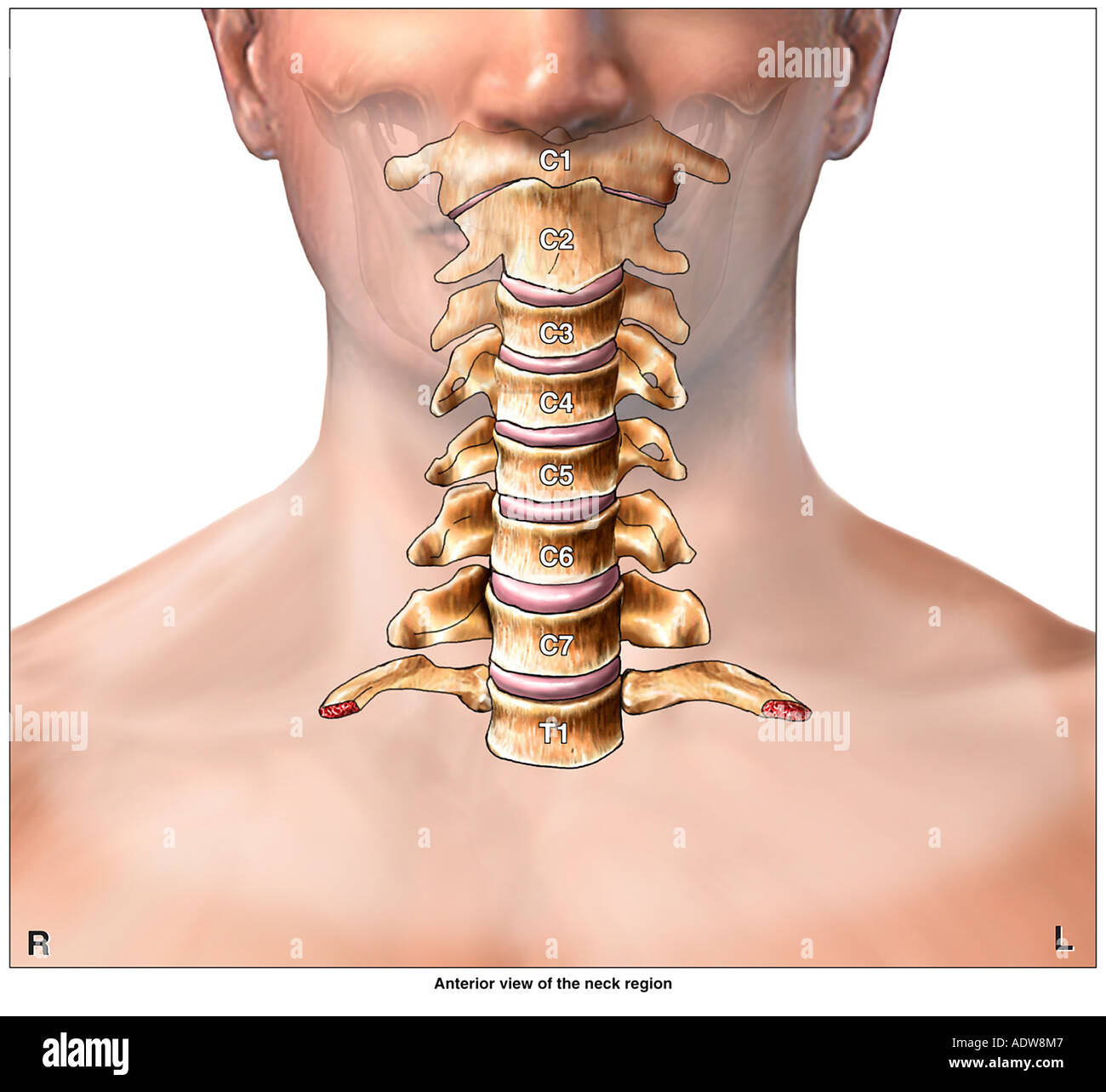
Clinical Definition and Pathophysiology of Cervical Adenopathies
Cervical adenopathies refer to the pathological enlargement of lymph nodes in the neck, a common clinical finding with diverse etiologies ranging from benign reactive processes to malignant transformations. The cervical lymph node network is organized into distinct anatomical chains that reflect both superficial and deep lymphatic drainage territories, necessitating a systematic approach to localization and differential diagnosis. Understanding the underlying mechanisms—whether infectious, inflammatory, or neoplastic—is critical for accurate diagnosis, as these processes trigger distinct histological and clinical presentations.
The cervical lymph node system is anatomically divided into superficial and deep groups, each with specific drainage territories that correlate with underlying pathologies. Superficial nodes include the occipital, posterior auricular, anterior auricular, submandibular, submental, and superficial cervical chains, while deep nodes comprise the jugulodigastric, jugulo-omohyoid, spinal accessory, supraclavicular, and paratracheal groups. Each chain drains distinct anatomical regions, with deep cervical nodes often serving as conduits for metastatic spread from head and neck malignancies.
Anatomical Distribution and Drainage Territories of Cervical Lymph Nodes
The cervical lymph node chains follow a hierarchical organization based on their anatomical position and functional drainage. Superficial nodes primarily drain cutaneous and mucosal surfaces of the head and neck, while deep nodes receive lymphatic flow from deeper structures, including the thyroid, parotid glands, and upper respiratory tract.Key Drainage Territories by Chain:The deep cervical chain, particularly the jugulodigastric nodes, is frequently involved in infectious processes such as tonsillitis or pharyngitis, while supraclavicular adenopathy raises suspicion for metastatic disease (e.g., lymphoma, breast, or lung cancer). Knowledge of these territories aids in localizing the primary pathology based on node involvement.
Occipital and Posterior Auricular Nodes: Drain the scalp, occipital region, and posterior auricular area. Anterior Auricular and Parotid Nodes: Receive lymph from the external ear, temporal region, and parotid gland. Submandibular and Submental Nodes: Drain the oral cavity, lips, nasal cavity, and anterior tongue. Superficial Cervical Nodes: Collect lymph from the skin of the neck and superficial structures. Deep Cervical Nodes (Jugulodigastric, Jugulo-Omohyoid): Primarily drain the nasopharynx, oropharynx, and upper aerodigestive tract. Supraclavicular Nodes: Serve as a critical sentinel for metastatic spread from thoracic and abdominal malignancies (e.g., Virchow’s node in gastric cancer). Spinal Accessory Nodes: Drain the posterior scalp and deep cervical structures.
Pathophysiological Mechanisms of Lymph Node Enlargement
Lymph node enlargement (lymphadenopathy) results from three primary mechanisms: reactive hyperplasia, granulomatous inflammation, and neoplastic infiltration. Each mechanism triggers distinct histological changes and clinical presentations, requiring careful evaluation.Core Pathophysiological Triggers:The immune response to antigens involves T-cell and B-cell activation, leading to follicular hyperplasia in reactive nodes. In contrast, neoplastic processes replace normal nodal architecture with malignant cells, often accompanied by capsular disruption and extracapsular extension. Granulomatous inflammation, characterized by epithelioid macrophages and multinucleated giant cells, is typically seen in infections (e.g., tuberculosis) or autoimmune disorders.
Infectious: Bacterial (e.g., Streptococcus, Mycobacterium tuberculosis), viral (e.g., EBV, CMV), or fungal agents induce lymph node hyperplasia via antigen presentation and immune activation. Inflammatory: Autoimmune conditions (e.g., rheumatoid arthritis, systemic lupus erythematosus) or non-infectious granulomatous diseases (e.g., sarcoidosis) provoke lymphoid proliferation. Neoplastic: Primary lymphomas (e.g., Hodgkin’s lymphoma) or metastatic spread (e.g., squamous cell carcinoma) disrupt nodal architecture, leading to pathological enlargement.
Differentiating Reactive Hyperplasia from Pathological Adenopathy
Histological examination remains the gold standard for distinguishing benign reactive changes from malignant or granulomatous pathologies. Key features include follicular architecture, cellular composition, and presence of necrosis or atypical cells.Histological Differentiation Criteria:Immunohistochemical staining (e.g., CD20 for B-cells, CD3 for T-cells, CD15/CD30 for Hodgkin’s lymphoma) further refines diagnosis. Flow cytometry is essential for identifying clonal populations in suspected lymphomas.
Reactive Hyperplasia: Follicular hyperplasia: Expanded germinal centers with polarized B-cell zones. Paracortical hyperplasia: Predominance of T-cells in response to viral infections. Preserved nodal architecture: Intact capsule and sinus structure. Granulomatous Inflammation: Epithelioid macrophages: Clustered with or without caseous necrosis (e.g., tuberculosis). Langerhans giant cells: Multinucleated cells in chronic granulomas. Lymphocyte cuffing: Surrounding inflammatory infiltrate. Neoplastic Adenopathy: Disrupted architecture: Loss of normal nodal zones. Atypical cells: Large, irregular nuclei with high mitotic activity (e.g., Reed-Sternberg cells in Hodgkin’s lymphoma). Extracapsular spread: Invasion into adjacent tissues.
Comparative Table: Types of Cervical Adenopathy
The following table summarizes the key features of common cervical adenopathy etiologies for rapid clinical reference.| Type of Adenopathy | Etiology | Histological Features | Key Clinical Signs |
|---|---|---|---|
| Reactive Lymphadenitis | Bacterial/viral infections (e.g., streptococcal pharyngitis, mononucleosis), autoimmune disorders | Follicular hyperplasia, paracortical expansion, preserved capsule | Fever, pharyngotonsillar erythema, tender nodes, self-limited course |
| Granulomatous Lymphadenitis | Tuberculosis, fungal infections (e.g., histoplasmosis), sarcoidosis | Epithelioid macrophages, caseous necrosis, granulomas with giant cells | Painless, rubbery nodes; systemic symptoms (weight loss, night sweats) |
| Lymphoma (Hodgkin’s) | Malignant proliferation of Reed-Sternberg cells (B-cell origin) | Mixed cellularity with lacunar cells, fibrosis, nodal effacement | Painless cervical/supraclavicular adenopathy, B symptoms (fever, weight loss) |
| Metastatic Adenopathy | Squamous cell carcinoma (head/neck), melanoma, breast cancer | Disrupted architecture, atypical epithelial cells, extracellular mucin (if mucinous carcinoma) | Hard, fixed nodes; associated primary tumor signs (e.g., oral ulcer, skin lesion) |
| Cat-Scratch Disease | Bartonella henselae infection (scratch/bit from infected cat) | Necrotizing granulomas, stellate abscesses, vascular proliferation | Regional lymphadenopathy, history of cat exposure, self-limited |

Etiological Classification and Differential Diagnosis of Cervical Adenopathies
Cervical adenopathy presents a broad spectrum of etiologies, ranging from benign inflammatory responses to malignant transformations. The differential diagnosis requires a systematic approach integrating patient demographics, geographic exposure, clinical presentation, and ancillary testing. Age-specific infectious agents, neoplastic risk factors, and autoimmune markers significantly influence diagnostic precision. This section categorizes cervical adenopathy by etiology, emphasizing infectious causes stratified by age, neoplastic classifications with prognostic indicators, and autoimmune/inflammatory mimics with diagnostic criteria.Infectious Etiologies Stratified by Age and Geographic Prevalence
Infectious agents account for a substantial proportion of cervical adenopathies, with pathogen distribution varying markedly across age groups and regions. Understanding these patterns facilitates targeted diagnostic workups and empirical therapies. Geographic prevalence, immune status, and vaccination histories further refine the likelihood of specific pathogens.Children and Adolescents (0–18 years)
In this age group, viral and bacterial infections dominate, often presenting with acute or subacute cervical lymphadenopathy.
- Viral Causes
- Epstein-Barr virus (EBV): Causes infectious mononucleosis, characterized by generalized lymphadenopathy, pharyngitis, and fatigue. Serology (heterophile antibodies, anti-VCA IgM) confirms diagnosis. Peak incidence in adolescents (15–24 years).
- Human herpesvirus 6 (HHV-6): Associated with exanthem subitum (roseola) in infants and may present with cervical adenopathy in immunocompromised children.
- Cytomegalovirus (CMV): Less common than EBV but may cause cervical adenopathy in immunocompromised or post-transplant pediatric patients.
- Human immunodeficiency virus (HIV): Primary infection (acute retroviral syndrome) may present with cervical adenopathy, particularly in high-risk populations.
- Group A Streptococcus (GAS): Causes suppurative cervical lymphadenitis (e.g., "hot potato" lymphadenitis) in children aged 2–5 years, often secondary to pharyngitis or skin infections. Streptococcus pyogenes is the predominant pathogen.
Infectious cervical adenopathy in adults often reflects reactivation of latent infections, occupational exposures, or sexually transmitted diseases (STDs).
- Viral Causes
- EBV and CMV: Reactivation in immunocompromised adults (e.g., post-transplant, HIV/AIDS) may present with persistent cervical adenopathy.
- HIV: Chronic generalized lymphadenopathy (CGL) is a hallmark of early HIV infection, with bilateral cervical lymphadenopathy lasting >3 months.
- Human T-lymphotropic virus type 1 (HTLV-1): Endemic in Japan, the Caribbean, and South America; associated with adult T-cell leukemia/lymphoma (ATLL) and tropical spastic paraparesis.
- Mycobacterium tuberculosis: The most common cause of chronic cervical adenopathy in adults, particularly in high-burden regions (e.g., sub-Saharan Africa, Southeast Asia). Caseating granulomas on fine-needle aspiration (FNA) or excisional biopsy are diagnostic.
- Toxoplasma gondii: Reactivation in immunocompromised adults (e.g., HIV/AIDS with CD4 <100 cells/µL) causes necrotizing cervical adenopathy. Serology (IgG/IgM) or PCR of lymph node tissue confirms infection.
Opportunistic infections dominate in this population, with pathogens reflecting the underlying immunodeficiency.
- Viral Causes
- CMV and EBV: Reactivation in post-transplant or HIV/AIDS patients leads to persistent cervical adenopathy.
- HHV-8 (Kaposi’s sarcoma-associated herpesvirus): Causes cervical lymphadenopathy in advanced HIV/AIDS or post-transplant patients.
- Nocardia spp.: Opportunistic infection in immunocompromised hosts, presenting with cervical adenopathy and pulmonary involvement.
- Histoplasma capsulatum: Cervical adenopathy in endemic regions (e.g., Ohio/Mississippi River valleys) or disseminated disease in immunocompromised patients.
Neoplastic Etiologies and Risk Factors
Neoplastic cervical adenopathy encompasses primary lymphomas, metastatic disease, and rare hematologic malignancies. B-symptoms (fever, night sweats, weight loss) and specific risk factors guide diagnostic suspicion.Lymphomas
Primary lymphomas of the cervical nodes are classified into Hodgkin lymphoma (HL) and non-Hodgkin lymphoma (NHL), with distinct epidemiologic and clinical features.
- Hodgkin Lymphoma (HL)
- Epidemiology: Bimodal age distribution (peak incidence in 20s and >55 years). Associated with Epstein-Barr virus (EBV) in mixed cellularity and nodular sclerosis subtypes.
- Risk Factors:
- EBV infection (seropositivity for anti-EBV VCA IgG).
- Family history or genetic predisposition (e.g., TNFRSF14 mutations).
- Immunosuppression (e.g., HIV, post-transplant).
- Clinical Features:
Cervical lymphadenopathy (often painless, rubbery, and progressive) with B-symptoms in ~30% of cases. Mediastinal involvement is common in nodular sclerosis subtype.
- Diagnostic Workup:
- Excisional biopsy with Reed-Sternberg cells (owl-eyed macrophages) on histology.
- Immunohistochemistry (CD15+, CD30+, CD20-).
- PET-CT to stage disease (Ann Arbor classification).
- Epidemiology: More common than HL, with incidence increasing with age. Subtypes include diffuse large B-cell lymphoma (DLBCL), follicular lymphoma (FL), and mantle
- Shotty nodes: Multiple, small (<1 cm), discrete, and mobile nodes commonly seen in viral infections (e.g., EBV, CMV) or autoimmune conditions (e.g., sarcoidosis). They lack tenderness unless superimposed cellulitis is present.
- Matted nodes: Confluent, irregularly shaped masses with loss of discrete borders, indicative of suppurative lymphadenitis (e.g., bacterial abscesses) or metastatic disease with extranodal extension.
- Echogenicity: Hypoechoic (dark) nodes with loss of echogenic hilum are suspicious for malignancy, while hyperechoic nodes with preserved hilum suggest benign etiologies (e.g., reactive lymphadenopathy).
- Border Regularity: Well-defined, smooth borders favor benign processes, whereas irregular or microlobulated borders are associated with malignancy.
- Central Hilar Echo: Absence of a central hilar echo (the "hilar sign") correlates with a 90% likelihood of malignancy in some studies.
- Hypervascularity: Seen in inflammatory or infectious adenopathy (e.g., bacterial lymphadenitis, Kikuchi disease), where increased blood flow reflects reactive hyperplasia.
- Hypovascularity: Characteristic of malignant nodes, where neovascularization is often chaotic and peripheral (e.g., peripheral vascularity in lymphomas or metastases).
- Avascular Nodes: Rare but highly suggestive of necrosis (e.g., abscesses or advanced tumors).
- Measurement Criteria: Nodes >1 cm in the short axis are considered abnormal, though size alone lacks specificity. Central necrosis or contrast enhancement patterns refine suspicion.
- Contrast Enhancement:
- Ring enhancement: Pathognomonic for abscesses (e.g., bacterial or mycobacterial lymphadenitis), where peripheral vascularity reflects the inflammatory capsule.
- Homogeneous enhancement: Seen in lymphomas, metastases, and reactive nodes, though homogeneous uptake in malignancies is often less intense than in infections.
- Necrosis: Low-attenuation centers (<20 HU) on contrast-enhanced CT suggest necrosis, common in advanced tumors (e.g., squamous cell carcinoma) or fungal infections (e.g., histoplasmosis).
- Extranodal Extension: Irregular infiltration beyond nodal borders into adjacent fat or muscles is a red flag for malignancy (e.g., head and neck squamous cell carcinoma).
- T1/T2 Signal Characteristics:
- T1-hypointense, T2-hyperintense: Typical of lymphomas and some metastases.
- T1-hyperintense: Rare but seen in fat-containing lesions (e.g., lipomatous lymph nodes in HIV-associated lymphadenopathy).
- Diffusion-Weighted Imaging (DWI): Restricted diffusion (high signal on DWI, low on apparent diffusion coefficient [ADC] maps) is highly suggestive of malignancy, as seen in lymphomas and aggressive metastases.
- Contrast Enhancement:
- Lymphomas: Homogeneous enhancement with possible central necrosis.
- Metastases: Heterogeneous enhancement due to tumor heterogeneity.
- Granulomatous Disease: Low-grade enhancement with possible rim enhancement (e.g., tuberculosis).
- CT: Poor contrast resolution for small nodes (<5 mm) and susceptibility to motion artifacts.
- MRI: Longer scan times, higher cost, and contraindications (e.g., pacemakers, claustrophobia).
- Fixed or matted nodes with loss of mobility, indicating potential extranodal extension or advanced malignancy.
- Asymmetric enlargement (>1 cm in short axis) with rapid growth (doubling in size within 2–4 weeks), suggesting aggressive pathology (e.g., lymphoma, squamous cell carcinoma).
- Unexplained weight loss (>10% body weight over 6 months) or systemic symptoms (fever, night sweats, fatigue), which may indicate systemic lymphoma or metastatic disease.
- Overlying skin changes such as ulceration, erythema nodosum, or violaceous plaques, which may signal cutaneous lymphoma (e.g., mycosis fungoides) or disseminated infection (e.g., fungal adenopathy in immunocompromised patients).
- Contralateral cranial nerve palsies (e.g., CN XII, CN XI) or hoarseness, suggesting nodal compression or an occult primary tumor in the head and neck region.
- Prior history of malignancy (e.g., head and neck, breast, or lung cancer) with new-onset cervical lymphadenopathy, increasing suspicion for recurrence or metastasis.
- Immunocompromised status (HIV, post-transplant, chemotherapy), where atypical infections (e.g., Mycobacterium avium, Pneumocystis jirovecii) or opportunistic malignancies (e.g., Kaposi sarcoma) may present with atypical lymphaden
- First-line: Penicillin V (50 mg/kg/day divided q6h, max 2 g/day) or amoxicillin (50 mg/kg/day divided q8h) for 10–14 days (GAS coverage).
- MRSA-suspected or penicillin-allergic: Clindamycin (30–40 mg/kg/day divided q6–8h, max 1.8 g/day) or trimethoprim-sulfamethoxazole (TMP-SMX) (8–10 mg/kg/day TMP component, divided q12h) for 7–10 days.
- Severe cases (abscess, systemic symptoms): IV nafcillin/oxacillin (100–200 mg/kg/day divided q4–6h) or vancomycin (60 mg/kg/day divided q6–8h) for 2–3 weeks, followed by oral suppression (e.g., clindamycin).

Diagnostic Workflow: From History to Advanced Imaging in Cervical Adenopathy
The evaluation of cervical adenopathy follows a structured, evidence-based approach that integrates clinical history, meticulous physical examination, and advanced imaging to distinguish benign from malignant or infectious etiologies. A systematic workflow ensures timely identification of red flags while minimizing unnecessary interventions. This section outlines the step-by-step diagnostic process, emphasizing palpation techniques, imaging modalities, and key radiographic features that guide clinical decision-making.Physical Examination: Palpation Techniques and Node Characteristics
The physical examination of cervical lymphadenopathy begins with a detailed inspection of the neck for asymmetry, erythema, or visible masses, followed by bimanual palpation to assess node mobility, consistency, and tenderness. Proper technique involves using the fingertips to apply gentle, systematic pressure while comparing bilateral regions. Nodes are classified based on their mobility, size, and texture:- Mobility and Fixation:
Nodes freely mobile in all directions suggest reactive or inflammatory causes, whereas fixation to underlying structures (e.g., skin, fascia, or muscles) raises suspicion for malignancy or advanced infection. For example, a node fixed to the sternocleidomastoid muscle may indicate metastatic squamous cell carcinoma from an occult head and neck primary.
- Consistency and Texture:
Soft, discrete nodes (<1 cm) with normal overlying skin are often reactive, while firm or rubbery nodes warrant further evaluation. Hard, stony nodes with irregular borders are highly suggestive of malignancy. The terms "shotty" and "matted" describe distinct patterns:
- Tenderness and Overlying Skin Changes:
Acute tenderness with erythema suggests an infectious process (e.g., Streptococcus or Staphylococcus lymphadenitis), whereas painless nodes may reflect malignancy or granulomatous disease (e.g., tuberculosis, lymphoma). Overlying skin changes, such as ulceration or violaceous discoloration, may indicate underlying malignancy (e.g., cutaneous lymphoma) or disseminated fungal infections.
Role of Ultrasound in Cervical Adenopathy Evaluation
Ultrasound (US) is the first-line imaging modality for cervical lymphadenopathy due to its accessibility, lack of ionizing radiation, and ability to guide interventional procedures. Key features assessed include echogenicity, vascularity (via Doppler), and node morphology. The B-mode ultrasound evaluates:Doppler ultrasound assesses vascularity patterns, which differ significantly between infectious and neoplastic processes:
Ultrasound-guided fine-needle aspiration (FNA) is performed for nodes >1 cm or those with suspicious features. The procedure involves:
1. Preparation: Sterile technique, local anesthesia (if needed), and real-time US guidance to target the lesion.
2. Aspiration: Use of a 22–25-gauge needle with 10–20 passes to obtain cellular material for cytology.
3. Post-procedure: Immediate smear preparation (Diff-Quik or Papanicolaou stain) for rapid on-site evaluation (ROSE) to assess adequacy.
4. Follow-up: Cellular samples are sent for cytopathology, flow cytometry (for lymphomas), or microbiological cultures (if infection is suspected).
Limitations: US has operator dependency and may miss deep or retropharyngeal nodes. False negatives can occur with small or necrotic tumors.
Computed Tomography (CT) and Magnetic Resonance Imaging (MRI) in Cervical Adenopathy
When ultrasound findings are equivocal or nodes are inaccessible to palpation, cross-sectional imaging with CT or MRI provides detailed anatomical and contrast-enhanced information. Both modalities assess node size, shape, borders, and enhancement patterns, though MRI offers superior soft-tissue contrast.CT Findings:
MRI Findings:
Comparison of CT and MRI:
| Feature | CT | MRI |
|---|---|---|
| Soft-tissue resolution | Good (but limited vs. MRI) | Superior |
| Contrast use | Iodinated contrast (IV) | Gadolinium-based (IV) |
| Radiation exposure | Yes | No |
| Best for | Bone/airway assessment, rapid screening | Detailed nodal characterization, DWI |
Red Flags Requiring Immediate Referral
Certain clinical and radiographic features mandate urgent evaluation by an otolaryngologist, hematologist, or oncologist to rule out malignancy or life-threatening infections. The following red flags warrant expedited referral:Therapeutic Strategies by Etiology in Cervical Adenopathies
The management of cervical adenopathies varies significantly based on the underlying etiology, requiring tailored therapeutic approaches that balance efficacy, safety, and the risk of complications. Evidence-based protocols distinguish between bacterial, viral, neoplastic, and reactive causes, with treatment strategies ranging from empiric antibiotics to systemic chemotherapy. This section outlines structured therapeutic algorithms for each major etiology, emphasizing first-line interventions, follow-up criteria, and critical complications to monitor.
Bacterial Cervical Adenitis: Etiology-Specific Antibiotic Protocols
Bacterial cervical adenitis is most commonly caused by Streptococcus pyogenes (group A streptococcus, GAS) and Staphylococcus aureus, including methicillin-resistant S. aureus (MRSA). Treatment selection depends on bacterial susceptibility, local epidemiology, and clinical severity. Empiric therapy should cover both GAS and S. aureus, with adjustments based on culture results and response.Antibiotic Selection and Duration
For uncomplicated bacterial adenitis (e.g., localized pain, erythema, fluctuance without systemic toxicity):
Viral Cervical Adenopathy: Supportive Care and Antiviral Indications
Viral etiologies, such as Epstein-Barr virus (EBV), cytomegalovirus (CMV), adenovirus, and HIV, often present with generalized lymphadenopathy and self-limited courses. Management focuses on supportive care, symptom control, and monitoring for complications. Antiviral therapy is reserved for severe or immunocompromised patients.Supportive Measures
Antiviral Therapy IndicationsSymptom management: Fever/pain: Acetaminophen or ibuprofen (avoid NSAIDs in EBV if splenomegaly is present due to bleeding risk). Airway obstruction: Rare but requires urgent steroids (dexamethasone 0.6 mg/kg IV) or tracheostomy in cases of massive lymphadenopathy (e.g., EBV-associated angioedema). Dehydration: IV fluids for high fevers or poor oral intake. Monitoring: Hematologic parameters: CBC with differential (watch for leukopenia/thrombocytopenia in EBV/CMV). Liver function tests: Elevated transaminases in EBV/CMV (monitor for hepatitis). Airway patency: Flexible laryngoscopy if stridor or dysphagia develops.
Complications Requiring InterventionEBV (infectious mononucleosis): No antiviral indicated for immunocompetent patients; supportive care suffices. Steroids (prednisone 1–2 mg/kg/day) for <7 days in cases of severe airway compromise or hemolytic anemia. Ganciclovir/valganciclovir reserved for EBV-associated hemophagocytic lymphohistiocytosis (HLH) or severe immunosuppression. CMV: Ganciclovir (5 mg/kg IV q12h) or valganciclovir (900 mg PO bid) for 2–3 weeks in immunocompromised patients (e.g., HIV with CD4 <50 cells/μL or post-transplant). Foscarnet for ganciclovir-resistant strains. HIV-related lymphadenopathy: ART initiation if CD4 <350 cells/μL; lymphadenopathy may resolve with viral suppression. Prophylaxis: Trimethoprim-sulfamethoxazole for Pneumocystis jirovecii if CD4 <200 cells/μL.
Neoplastic Cervical Adenopathy: Staging and Treatment Algorithms
Neoplastic causes, including lymphomas and metastatic nodes, require multimodal therapy based on histologic subtype, staging, and performance status. Treatment algorithms are standardized for common entities such as Hodgkin lymphoma (HL), diffuse large B-cell lymphoma (DLBCL), and squamous cell carcinoma (SCC) metastases.Lymphoma Management
Staging Systems:Metastatic Neck Nodes (Squamous Cell Carcinoma)
Hodgkin lymphoma: Ann Arbor staging (I–IV) with PET-CT for metabolic activity. Non-Hodgkin lymphoma (NHL): Lugano classification (I–IV) with B symptoms (fever, night sweats, weight loss) guiding urgency. Treatment Protocols:
Hodgkin lymphoma (early-stage IA–IIA): ABVD (doxorubicin, bleomycin, vinblastine, dacarbazine) × 2 cycles followed by involved-node radiation (INRT 20–30 Gy). Alternative: AVD (without bleomycin) if pulmonary risk (e.g., COPD). Aggressive B-cell lymphoma (DLBCL): R-CHOP (rituximab, cyclophosphamide, doxorubicin, vincristine, prednisone) × 6–8 cycles for CD20+ disease. High-risk (e.g., MYC/BCL2 rearranged): DA-EPOCH-R or R-DHAP followed by autologous stem cell transplant (ASCT). Indolent lymphomas (e.g., follicular lymphoma): Watch-and-wait for asymptomatic patients; rituximab monotherapy or bendamustine-rituximab for symptomatic disease.
Treatment depends on primary site and nodal burden:Follow-Up Criteria for Neoplastic Adenopathy
Early-stage (N1, <3 cm, no extracapsular extension): Surgical excision (neck dissection) + adjuvant radiation (60–66 Gy) if high-risk features (perineural invasion, close margins). Advanced-stage (N2–N3 or extracapsular extension): Definitive chemoradiation (cisplatin 100 mg/m² q3w ×3 + 70 Gy) or induction chemotherapy (TPF: docetaxel, cisplatin, 5-FU) followed by surgery. Palliative cases (unresectable, symptomatic): Radiation (30 Gy in 10 fractions) or systemic therapy (cetuximab + platinum).
Comprehensive Treatment Decision Table
| Etiology |
|---|
| Histology | 5-Year OS (Localized) | 5-Year OS (Advanced) | Key Poor Prognostic Factors |
|---|---|---|---|
| Classical Hodgkin Lymphoma | 90% | 60–70% | Bulky mediastinal disease, B symptoms |
| Diffuse Large B-Cell Lymphoma | 70% | 30–40% | MYC/BCL2 double expressor, TP53 mutations |
| Squamous Cell Carcinoma (HNSCC) | 65% (N0) | 20–30% (N3) | Perineural invasion, HPV-negativity (p16-) |
| Papillary Thyroid Cancer | 95% (N1a) | 50% (N1b) | Extranodal extension, BRAF^V600E mutation |
Role of PET-CT in Staging and Restaging Cervical Adenopathy
Positron emission tomography-computed tomography (PET-CT) integrates metabolic activity with anatomical detail, improving accuracy in nodal staging and response assessment. However, interpretation requires awareness of false-positive and false-negative scenarios.Staging Applications
Restaging and Response Assessment
PET-CT in Specific Entities
The evaluation and management of cervical adenopathies exemplify the intersection of clinical acumen, diagnostic precision, and evidence-based therapy. By systematically assessing anatomical locations, etiologic triggers, and histological patterns, clinicians can navigate the complexities of this condition with greater confidence. The integration of advanced imaging, such as Doppler ultrasound and contrast-enhanced MRI, enhances diagnostic accuracy, while therapeutic algorithms tailored to infectious, inflammatory, or neoplastic causes optimize patient outcomes. Prognostic indicators and restaging tools, including PET-CT, further refine risk stratification and guide long-term surveillance. Ultimately, a multidisciplinary approach—spanning infectious disease specialists, oncologists, and radiologists—ensures comprehensive care for patients presenting with cervical lymphadenopathy, balancing early intervention with personalized treatment strategies.
Leave a Comment
Comments are moderated before appearing. The data you submit is processed according to the Privacy Policy of Ns2.